

For general education, not medical advice. This guide can’t diagnose you or replace care from your own clinician. If you’re worried about a spot or symptom, see a board-certified dermatologist.
Melanoma in situ is the earliest form of melanoma — "in situ" means it's still confined to the epidermis, the top layer of skin. It has not grown into the deeper layers where it could reach blood vessels or lymphatics, so while it remains in situ it cannot spread. That's why, despite the word "melanoma," the outlook is excellent: removing it completely is essentially curative.
Key points
- Melanoma in situ is Stage 0 — confined to the surface, with no ability to spread while it stays in situ.
- Removing it completely is essentially curative, usually with a minor outpatient excision.1
- It's a signal to watch your skin closely afterward, since one skin cancer raises the odds of another.
If you've been told you have melanoma in situ, this is the reassuring end of the melanoma spectrum — but it still deserves prompt, complete treatment and ongoing skin checks.
What is melanoma in situ?
Melanoma begins in melanocytes, the pigment-producing cells of the skin. In melanoma in situ, those abnormal cells are still contained within the epidermis. Left alone over time, some can progress to invasive melanoma, which is why it's treated rather than watched. A closely related subtype that arises on chronically sun-damaged skin — typically the face of older adults — is lentigo maligna, which behaves differently enough to have its own guide.
Signs to watch for
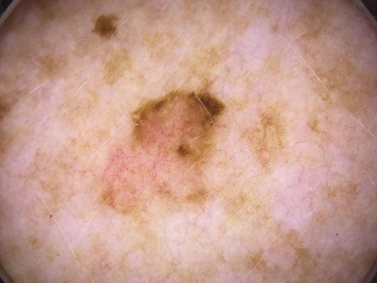
It usually looks like a new or changing flat spot or patch, often tan to brown to black, sometimes with uneven color or an irregular border — the ABCDEs (asymmetry, border, color, diameter, evolving). Many are found during a routine skin exam before they cause any symptom at all, which is the case for early detection.
How it's diagnosed
A suspicious spot is examined, often with a dermoscope, and confirmed with a skin biopsy — the only way to diagnose melanoma. The pathologist confirms the cells are confined to the epidermis (in situ) rather than invasive, which is what places it at Stage 0.
What treatment usually looks like
Because the abnormal cells sit only at the surface, the goal is simply to remove them completely. For most lesions that means surgical excision with a small margin of normal-looking skin, done in the office under local anesthesia — and that's typically the end of it.2 For lesions with hard-to-see borders, a margin-controlled (staged) approach may be used to confirm the edges are clear before closing.
Which approach your dermatologist recommends depends on the lesion's size, location, and how well-defined its borders are. There's no chemotherapy and no systemic treatment for in-situ disease — the work is local and, once margins are clear, done. Surgery is the standard, but the right plan is a shared decision with your doctor: for most people that means complete removal, while for a few — for example, someone with very limited life expectancy, or where an operation would carry more risk than a slow-growing surface lesion — careful monitoring can be a reasonable choice made together.3
Margins: why a little extra skin is removed
When the spot is removed, your doctor also takes a thin rim of normal-looking skin around it. That rim is called the margin. A clear margin means no abnormal cells are left at the edge.
Melanoma in situ can spread sideways under the skin, a little past what the eye can see. So a very small margin is not always enough. In one large study, a 5 mm margin cleared all the cells in about 8 out of 10 cases, while about 9 mm cleared nearly all of them.4 Your doctor checks the edges under a microscope. If any cells reach the edge, a little more skin is removed until the margin is clear. This is why complete removal works so well: once the edges are clear, the spot is gone.
Do I need scans or a lymph node test?
No. Because the cells stay in the top layer of skin, melanoma in situ cannot reach the lymph nodes or other organs. So body scans and a lymph node biopsy are not needed.2 Those tests are used for melanoma that has grown deeper. If a biopsy ever shows the melanoma went past "in situ," your care would change, and your doctor would talk you through the next steps.
Reducing your risk going forward
Having had a melanoma in situ raises your future risk, so the fundamentals matter more than ever: broad-spectrum sun protection, no tanning beds, monthly self-checks, and regular skin checks. For people with many moles, total body photography creates a baseline to track changes over time.
When to see a doctor
Have any new, changing, or unusual spot evaluated promptly — and keep your scheduled skin checks, since the most useful thing after one skin cancer is catching the next one early.
Worried about a spot?
SpotDoc offers full-body skin cancer screening in Brownwood Square, The Villages. No referral needed for most plans.
Frequently asked questions
Is melanoma in situ dangerous? While it stays in situ it cannot spread, and complete removal is essentially curative. It's treated promptly because, left alone, some lesions can progress to invasive melanoma over time.
Will I need anything besides surgery? Usually not. In-situ disease is treated locally — there's no role for chemotherapy or systemic therapy. Ongoing skin surveillance is the main follow-up.
How is this different from lentigo maligna? Lentigo maligna is a subtype of melanoma in situ on sun-damaged skin (usually the face) that spreads more widely under the surface, so it often needs wider or margin-controlled removal. See the lentigo maligna guide.
Can melanoma in situ spread to other parts of my body? Not while it stays "in situ." The cells sit in the top layer of skin, with no path to the blood or lymph nodes. The reason to treat it now is to remove it before it ever has the chance to grow deeper.
How big will the scar be? The area removed is a bit larger than the spot, because of the margin of normal skin around it. Most wounds close in a thin line. Spots on the face or larger lesions may need a more careful repair.
Can it come back after it's removed? When the whole lesion is removed with clear margins, it rarely comes back, about 1 in 100.1 More often, a "new" spot later is a separate skin cancer, which is why ongoing skin checks matter.
How often should I get my skin checked afterward? Most people have a full skin exam at least once a year for life, and sometimes more often at first. Your dermatologist sets the timing based on your skin, your moles, and your family history.
Questions to ask your dermatologist
- Were my margins clear, or do I need more skin removed?
- Was the melanoma only in situ, or did any of it go deeper?
- How often should I have my skin checked from now on?
- What changes should make me call the office sooner?
Reviewed by Tyler Long, DO. Last updated June 2026.
Update log
How this guide has changed since it was first published.
- August 18, 2026Guide published.
References
Every statement on this page is backed by the peer-reviewed sources below — each links to the original study. Numbers match the citations in the text and the grids above.
- Akhtar S, Bhat W, Magdum A, Stanley PRW. Surgical excision margins for melanoma in situ. J Plast Reconstr Aesthet Surg. 2013;67(3):320–323.View source
- Swetter SM, Tsao H, Bichakjian CK, et al. Guidelines of care for the management of primary cutaneous melanoma. J Am Acad Dermatol. 2019;80(1):208–250.View source
- National Comprehensive Cancer Network. NCCN Clinical Practice Guidelines in Oncology: Melanoma: Cutaneous. Version 2.2026. nccn.org. 2026.View source
- Kunishige JH, Brodland DG, Zitelli JA. Surgical margins for melanoma in situ. J Am Acad Dermatol. 2012;66(3):438–444.View source
- Kunishige JH, Doan L, Brodland DG, Zitelli JA. Comparison of surgical margins for lentigo maligna versus melanoma in situ. J Am Acad Dermatol. 2019;81(1):204–212.View source
- Nosrati A, Berliner JG, Goel S, et al. Outcomes of melanoma in situ treated with Mohs micrographic surgery compared with wide local excision. JAMA Dermatol. 2017;153(5):436–441.View source
- Felton S, Taylor RS, Srivastava D. Excision margins for melanoma in situ on the head and neck. Dermatol Surg. 2016;42(3):327–334.View source
- Beveridge J, Taher M, Zhu J, Mahmood MN, Salopek TG. Staged margin-controlled excision (SMEX) for lentigo maligna melanoma in situ. J Surg Oncol. 2018;118(1):144–149.View source
- Etzkorn JR, Sobanko JF, Elenitsas R, et al. Low recurrence rates for in situ and invasive melanomas using Mohs micrographic surgery with MART-1 immunostaining. J Am Acad Dermatol. 2015;72(5):840–850.View source
- Moyer JS, Rudy S, Boonstra PS, et al. Efficacy of staged excision with permanent section margin control for cutaneous head and neck melanoma. JAMA Dermatol. 2017;153(3):282–288.View source
- Clark GS, Pappas-Politis EC, Cherpelis BS, et al. Surgical management of melanoma in situ on chronically sun-damaged skin. Cancer Control. 2008;15(3):216–224.View source