
For general education, not medical advice. This guide can’t diagnose you or replace care from your own clinician. If you’re worried about a spot or symptom, see a board-certified dermatologist.
What Is Total Body Photography?
Total Body Photography (TBP) is a skin-imaging technology that captures a detailed, high-resolution record of the skin across most of the body in a single session. The images form a baseline "map" of your moles and other spots. At future visits, a dermatologist can compare new images against that baseline to see what is new, what has grown, and what has changed shape or color.
The principle behind TBP is simple but powerful: melanoma is often easiest to recognize not by how a single mole looks on one day, but by how a spot changes over time, or by a new lesion that does not match a person's other moles. A photographic baseline gives the clinician an objective reference instead of relying on memory or a patient's own recollection.
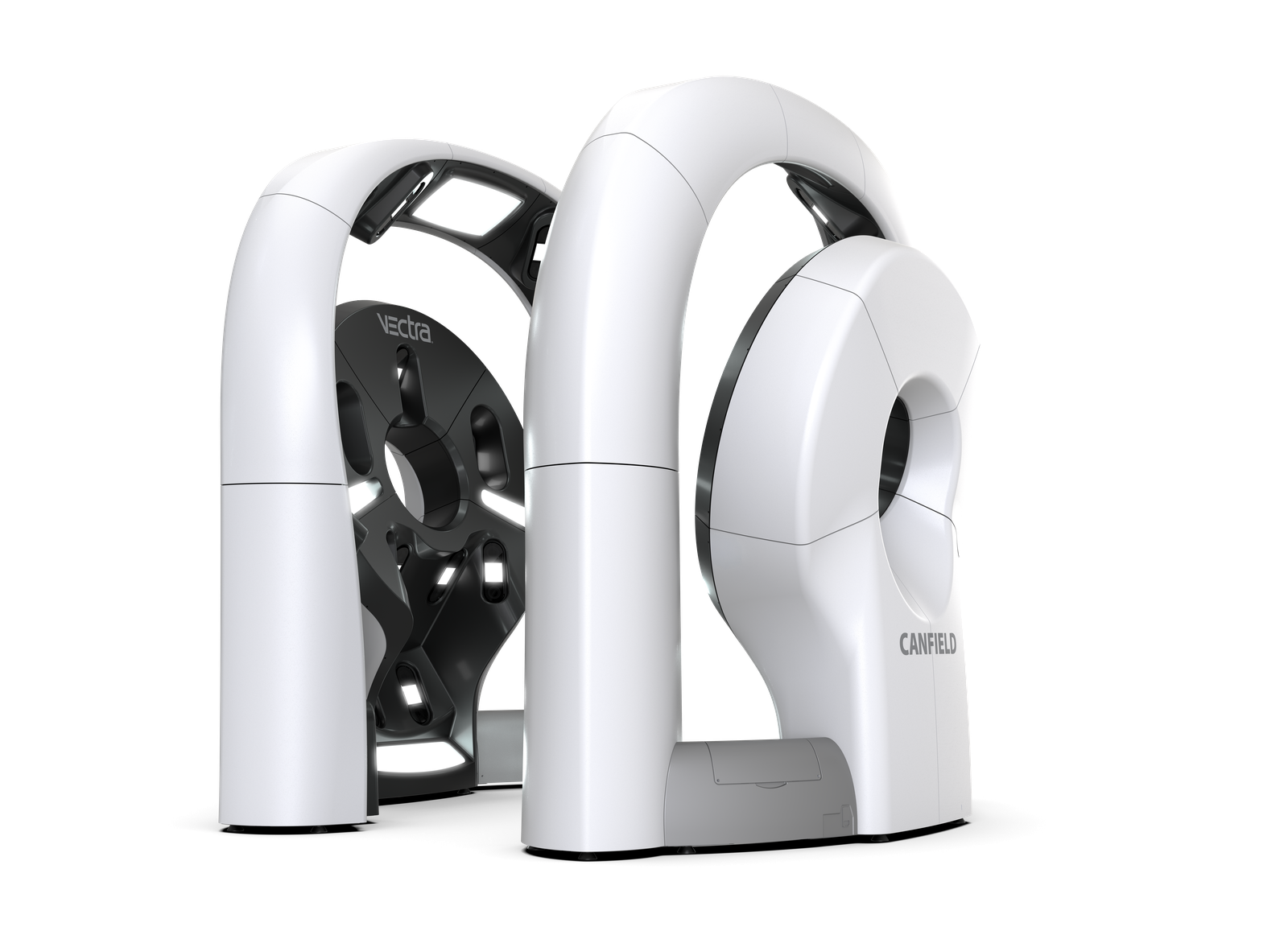
Modern systems extend this idea into three dimensions. A 3D Total Body Photography system uses an array of cameras positioned around the patient to capture the body surface almost simultaneously, then assembles the images into a navigable model. Individual lesions can be tagged and tracked, and close-up dermoscopic images of specific moles can be linked to their location on the map.
How It Works
A TBP session is non-invasive and involves no radiation or injections — it is photography. The general process is:
- Image capture: The patient stands in a series of standardized poses while a multi-camera system, or a clinician using a calibrated camera, photographs the skin surface. Capture of the full set of images typically takes only seconds to a few minutes.
- Map assembly: Software stitches the images into a complete view of the skin. In 3D systems, this becomes a rotatable avatar on which each lesion has a fixed location.
- Lesion documentation: Moles of interest can be marked, measured, and paired with magnified dermoscopic images for finer detail.
- Baseline storage: The images are stored securely as a reference point.
- Sequential comparison: At follow-up visits, new images are compared with the baseline. This side-by-side comparison over time is known as sequential digital dermoscopy imaging when applied to individual lesions, and it is what allows subtle change to be detected.
The technology is a documentation and monitoring tool. It supports the dermatologist's examination — it does not replace the clinical skin exam, the dermoscopic evaluation of individual lesions, or biopsy when a lesion is suspicious.
What It's Used For
TBP is primarily a surveillance tool for people at elevated risk of melanoma, where catching a new or changing lesion early matters most. Common uses include:
- Monitoring people with many moles, where examining and remembering each one is impractical
- Tracking atypical (dysplastic) moles that warrant watching rather than immediate removal
- Following patients with a personal or family history of melanoma, who carry higher lifetime risk
- Reducing unnecessary biopsies by giving the clinician evidence that a stable-looking mole has, in fact, been stable over time
- Documenting a baseline before starting long-term dermatologic follow-up
By making change easier to see, TBP can help focus attention — and biopsies — on the lesions most likely to matter, while leaving longstanding, stable moles alone.
Who Benefits Most
TBP is not necessary for everyone. For a person with few moles and no risk factors, a routine skin exam is usually sufficient. The technology delivers the most value for higher-risk individuals, including those with:
- A high total number of moles (a leading melanoma risk factor)
- Multiple atypical or dysplastic nevi
- A personal history of melanoma or non-melanoma skin cancer
- A family history of melanoma, including familial atypical multiple mole and melanoma (FAMMM) syndrome
- Fair skin, light eyes, a history of significant sun exposure or sunburns, or a weakened immune system
Whether TBP is appropriate for you is a clinical decision best made with a dermatologist who can weigh your individual risk factors.
What the Evidence Shows
Total Body Photography and sequential digital dermoscopy are established components of melanoma surveillance for high-risk patients, and international guidelines — including the European consensus-based interdisciplinary melanoma guidelines updated through 2024 — support their use to help detect early-stage melanoma and reduce biopsies of clearly benign lesions in this group. Long-term observational studies have documented that combining whole-body photography with digital dermoscopy can identify melanomas at an early, thin stage during follow-up of high-risk patients.
The evidence continues to evolve. Real-world and observational studies of combined total body photography and sequential digital dermoscopy report earlier identification of melanoma and fewer biopsies of clearly benign lesions among high-risk patients, while researchers continue to refine which patients benefit most and how often imaging should be repeated. TBP is most valuable when it is matched to the right patient and used as part of ongoing follow-up — it supports, rather than replaces, the dermatologist's examination and clinical judgment.
Research into computer-assisted and artificial-intelligence analysis of TBP images is active and ongoing, but such tools remain an area of study rather than a substitute for a dermatologist's evaluation. The interpretation of the images, and any decision to biopsy, rests with the clinician.
What to Expect at Your Appointment
A TBP session is straightforward and comfortable:
- Preparation: You will be asked to remove clothing down to undergarments so the skin can be imaged. Remove makeup, and it is helpful to avoid heavy lotions or self-tanner, which can obscure the skin. Long hair may be tied back.
- The imaging: You will stand in a few standardized positions while the images are captured. The capture itself is brief.
- The exam: A dermatologist reviews the images together with a hands-on skin examination and dermoscopy of any lesions of concern. The photographs supplement the exam; they do not replace it.
- Follow-up: Your images become the baseline for future comparison. Your dermatologist will recommend an interval for returning — in practice, photographic baselines are often repeated roughly every 1–2 years, with closer dermoscopic monitoring of specific lesions in between, adjusted to your risk.
Limitations & Considerations
TBP is a tool, not a diagnosis. A few practical points to keep in mind:
- It does not image every surface. Areas such as the scalp under hair, between toes, and mucous membranes still require direct examination.
- It does not detect cancer on its own. It documents the skin so a clinician can recognize change; the clinical judgment and any biopsy remain essential.
- More monitoring can mean more procedures. Heightened surveillance sometimes leads to additional biopsies or excisions of lesions that turn out to be benign — one reason careful patient selection matters.
- Coverage and cost vary. Total Body Photography may be offered as a self-pay service, and insurance coverage differs by plan and by individual circumstances. Ask your dermatology office about the cost and whether any portion may be billable to your insurance.
When to Consider Total Body Photography
If you have many moles, atypical moles, or a personal or family history of melanoma, ask your dermatologist whether Total Body Photography fits into your follow-up plan. It is most valuable as part of an ongoing surveillance relationship — the baseline today is what makes a change visible a year from now.
Regardless of whether TBP is right for you, the fundamentals of skin cancer detection still apply: perform regular skin self-exams, watch for the ABCDEs of melanoma (Asymmetry, Border irregularity, Color variation, Diameter over 6 mm, and Evolving), and see a dermatologist promptly about any spot that is new, changing, or simply does not look like your other moles.
References
- European consensus-based interdisciplinary guideline for melanoma. Part 1: Diagnostics — Update 2024. European Journal of Cancer. 2024. sciencedirect.com
- Salerni G, Carrera C, Lovatto L, et al. Characterization of 1152 lesions excised over 10 years using total-body photography and digital dermatoscopy in the surveillance of patients at high risk for melanoma. Journal of the American Academy of Dermatology. 2012;67(5):836–845. sciencedirect.com
- Clinical outcomes of 3D-total body photography and digital dermoscopy for surveillance of high-risk melanoma patients: a prospective longitudinal observational study. European Journal of Cancer. 2025. sciencedirect.com
- Three-Dimensional Total Body Photography, Digital Dermoscopy, and in vivo Reflectance Confocal Microscopy for Follow-Up Assessments of High-Risk Patients for Melanoma: A Prospective, Controlled Study. Dermatology. 2024;240(5–6):803–814. karger.com
- Melanoma surveillance for high-risk patients via telemedicine: examination of real-world data from an integrated store-and-forward total body photography and dermoscopy service. Journal of the American Academy of Dermatology. 2021. jaad.org
- The importance of total-body photography and sequential digital dermatoscopy for monitoring patients at increased melanoma risk. pmc.ncbi.nlm.nih.gov
- Clinical Perspective of 3D Total Body Photography for Early Detection and Screening of Melanoma. pmc.ncbi.nlm.nih.gov