# SpotDoc — The Skin Cancer Company
> SpotDoc — "The Skin Cancer Company" — is a skin-cancer screening company building a national network dedicated to finding skin cancer earlier. SpotDoc pairs SpotCheck 360° total body photography (full-body imaging on a Canfield Vectra WB360) with board-certified dermatologist review to map the skin and track changes over time. It operates through three models: Flagship Clinics owned and operated by SpotDoc, Partnership Practices that bring SpotDoc imaging into established dermatology groups, and Concierge Services at premium health centers. Its mission is to make early skin cancer detection more accessible across the country.
Network:
- Flagship clinic: The Villages, Florida — led by Tyler Long, DO (board-certified dermatologist) with Kim Nguyen, NP-C
- Partnership Practices: New Jersey (Red Bank and Ramsey)
- Concierge Services: New York City (Elitra Health)
- Expanding to new markets across the United States
How SpotCheck 360° works: a Canfield Vectra WB360 captures 92 photographs in about a second to build a full-body map of the skin; at each visit the new images are compared to the prior baseline so the team evaluates what is changing over time.
## About SpotDoc
SpotDoc — "The Skin Cancer Company" — is a skin-cancer screening company building a national network with one focus: finding skin cancer earlier. Its core service, SpotCheck 360°, uses total body photography on a Canfield Vectra WB360 to capture 92 photographs in about a second and build a full-body map of the skin; at each visit the new images are compared to the prior baseline so the clinical team evaluates what is changing over time rather than relying on memory. Every imaging study is reviewed by a board-certified dermatologist.
SpotDoc delivers care through three models:
- Flagship Clinics — owned and operated by SpotDoc, offering screening, evaluation, and treatment under one roof. The flagship is in The Villages, Florida.
- Partnership Practices — established dermatology groups that integrate SpotDoc imaging into their services: Navesink Dermatology (formerly Dermatology and Skin Cancer Center) in Red Bank, NJ, and Skin and Laser Center of NJ in Ramsey, NJ.
- Concierge Services — SpotDoc screening offered within premium health-assessment centers (currently Elitra Health in New York City).
SpotDoc is expanding to new markets across the United States, with a mission to make early skin cancer detection more accessible.
## Flagship clinic — The Villages, Florida
The Villages flagship is a physician-led clinic where skin cancer is the entire focus. Patients are seen by Tyler Long, DO, a board-certified dermatologist, and Kim Nguyen, NP-C. It houses the only Canfield Vectra WB360 within 280 miles.
What to expect: a typical visit runs about 45 minutes: a brief history, the total body photography (a ring of 92 cameras captures the entire skin surface in about a second), and a clinical exam with the team, who review any areas of concern and discuss next steps. Many treatments are done in-office.
Practical details: Medicare and most major commercial plans are accepted for the clinical exam, including Aetna, Cigna, Humana, UnitedHealthcare, and Blue Cross Blue Shield; no referral is needed for most plans (some Medicare Advantage HMOs, such as Humana Medicare Advantage, require one). The total body photography itself is currently an out-of-pocket charge, quoted before booking. Medicare Advantage plans are often out of network; coverage is verified before the visit. Located at 3614 Kiessel Road, Brownwood Square, The Villages, FL 32163. Book online or call (352) 914-3451.
## Frequently asked questions
### Insurance
**Is a screening covered by insurance?**
Partly, and the split is worth understanding. The clinical exam is typically covered the way any dermatology visit is. The total body photography itself is not currently covered by insurance, Medicare included; it is an out-of-pocket charge, quoted up front so there are no surprises. The Villages, FL (flagship clinic): Most major plans accepted for the exam. We verify your plan and quote the photography charge before your visit. New Jersey (partner practices): The partner practices bill through their own offices; verify coverage with them directly. New York (concierge program): Typically self-pay, as part of a comprehensive health assessment.
**Do you take Medicare, including Medicare Advantage?**
Traditional Medicare, yes: the flagship clinic and most partner practices accept it, and it generally covers medically necessary skin exams, such as when you have a history of skin cancer, notable risk factors, or a spot of concern. Medicare Advantage is plan by plan and often out of network, so call with your card handy and we will check yours before you book. Either way, the total body photography is a separate out-of-pocket charge, quoted in the same call.
**Which insurance plans do you accept?**
It depends on where you are seen, and in every case insurance applies to the clinical exam; the total body photography is an out-of-pocket charge. The Villages, FL (flagship clinic): Medicare and most major plans, including Aetna, Cigna, Humana, UnitedHealthcare, Blue Cross Blue Shield, Sunshine and WellCare, and MultiPlan. When in doubt, call with your card handy and we will check. New Jersey (partner practices): Each partner practice bills through its own office; verify your plan with them directly. New York (concierge program): Typically self-pay.
**What does it cost if I'm paying out of pocket?**
Self-pay patients are welcome, and pricing is straightforward but varies by location and visit type, so we quote it rather than publish numbers that drift out of date. The Villages, FL (flagship clinic): Call (352) 914-3451; the front desk gives current rates before you book, and payment plans can be discussed. New Jersey (partner practices): Partner practices set their own rates; call them directly. New York (concierge program): The concierge assessment is priced by Elitra Health; contact them directly.
### Appointments
**How do I schedule an appointment?**
It depends on your location. The Villages, FL (flagship clinic): Book online through our Klara scheduling system, or call (352) 914-3451. New Jersey (partner practices): Contact Navesink Dermatology in Red Bank or the Skin and Laser Center of NJ in Ramsey directly. New York (concierge program): Appointments are arranged through Elitra Health.
**How soon can I be seen?**
Availability changes week to week, so the honest answer lives in the online scheduler, which shows real open times at the flagship clinic in The Villages. If nothing online works, call (352) 914-3451 and the front desk will look for options. Partner practices in New Jersey manage their own calendars, so call them directly.
**How long does an appointment take?**
The scan itself takes about a second; the rest is your medical history and the head-to-toe exam with your clinician. The Villages, FL (flagship clinic): Plan on about 45 minutes for a full screening with total body photography. Follow-up visits are often shorter. New Jersey (partner practices): Visit lengths vary by practice; ask when you book. New York (concierge program): Concierge visits run longer because the screening is part of a broader health assessment.
**What should I wear to my appointment?**
Whatever is easy to change out of. You will wear a gown for the exam and imaging, the same as a regular dermatology visit. Skip makeup, heavy lotions, self-tanner, and sunscreen that day, since they interfere with image quality, and wear your hair so the scalp can be checked.
**Do I have to undress completely?**
You keep your underwear on. For the photography, the cameras need to see your skin, so you will be in minimal clothing for about a second in a private booth. For the hands-on exam you wear a gown, with one area uncovered at a time. A chaperone is available if you would like one; just ask.
**Can my spouse or a friend come with me?**
Yes. A companion is welcome in the consultation, and can step out during the photography and exam if you prefer privacy. Many patients bring their spouse; a second set of ears helps.
**What happens during a visit?**
Your clinician starts with a short history and any spots on your mind. You change into a gown and step into the imaging booth, where 92 cameras photograph your entire skin surface in about a second. Then comes the hands-on part: a head-to-toe exam, with the new images available for comparison against your baseline. You discuss anything of note together, and if a spot needs a biopsy, that can usually happen in the same visit.
**What happens if something concerning is found?**
You will know before you leave. Depending on the spot, your clinician may monitor it with closer follow-up, take a biopsy, or recommend treatment. At the flagship clinic, most diagnosis and treatment happens in the same office. Partner practices offer their own treatment options, and concierge patients receive referrals to the right specialists.
**How do I get my results?**
The screening itself has no waiting: your clinician goes over what they see during the visit. If a biopsy is taken, the sample goes to a pathology lab and we call you with the result as soon as it returns, whatever the answer is.
### Technology
**What is total body photography?**
A baseline record of your entire skin surface, captured as high-resolution images in about a second. It turns your skin into something that can be compared over time: at each visit, new images sit next to the old ones, and change becomes visible. Our clinical guide to total body photography covers it in depth.
**How does the imaging system work?**
You stand in a private booth and a ring of 92 cameras photographs your entire skin surface in about a second. Nothing touches you and there is no radiation; the system, a Canfield VECTRA WB360, uses ordinary visible light, like any camera. The images become a high-resolution map of your skin that serves as a baseline at future visits.
**Is this the same as mole mapping?**
It is the same idea, done comprehensively. Traditional mole mapping photographs selected moles one at a time. Total body photography captures the entire skin surface at once, so nothing depends on choosing the right spots to track in advance.
**Is the imaging process safe?**
Yes. Visible light only, no radiation, nothing invasive, no known side effects. It is safe at any age, during pregnancy, and with pacemakers or other implanted devices.
**What role does AI play in my screening?**
A supporting one. Software compares new images against your baseline and flags spots that look new or changed, and AI-assisted lesion review gives your clinician a second set of eyes. Every assessment and every recommendation comes from your clinician, not from a computer. We evaluate what is changing, not what no one can remember.
**Does the scan tell me whether a spot is cancer?**
No, and this is worth being clear about. The photography documents your skin and makes change visible over time; it does not diagnose anything. Diagnosis comes from your clinician's exam and, when a spot warrants it, a biopsy read by a pathologist. Think of the images as the map, not the verdict.
**Are there places the cameras can't see?**
Yes. The cameras cannot see through hair to the scalp, the bottoms of your feet while you stand, or deep skin folds such as the groin. That is why the visit is never photography alone: your clinician examines your scalp and feet directly at every screening, and examines the groin area on request. If you want it included, just say so.
**What does a screening look for?**
Your clinician examines you for all common skin cancers: melanoma, basal cell carcinoma, and squamous cell carcinoma, along with precancerous spots like actinic keratoses and atypical moles. The imaging supports that exam by making change over time visible.
**How do you track changes in my skin over time?**
Each visit adds a new set of images to your record, compared side by side against your baseline. A mole that has grown, or a spot that was not there last year, stands out. Anything flagged gets examined directly by your clinician.
**Can I get copies of my images?**
Yes. They are part of your medical record, and you have a right to your records. Ask at the office and we will arrange it.
**Is my data secure and private?**
Images and personal information are encrypted and stored in compliance with HIPAA. Only the clinicians involved in your care can access them, and we never share your information without your consent.
### General
**What is SpotDoc?**
SpotDoc is a dermatology practice built around one thing: skin cancer. Screening with total body photography, clinical exams, biopsies, and treatment. The flagship clinic is in The Villages, Florida, led by Tyler Long, DO, a board-certified dermatologist. Partner practices in New Jersey and a concierge program in New York carry the same screening approach.
**What makes a SpotDoc screening different?**
Most skin checks rely on the clinician's memory of your skin. Ours don't. Every screening pairs a head-to-toe exam with total body photography, so each visit is compared against a baseline map of your skin rather than against recollection. And because skin cancer is all we do, detection and treatment happen under one roof at the flagship clinic.
**Who will I see at my visit?**
At the flagship clinic in The Villages, the team is Tyler Long, DO, a board-certified dermatologist, and Kim Nguyen, NP-C. Visits are with the team, and because skin cancer is the entire practice, you are seen by clinicians who look at this all day. At the New Jersey partner practices, you are seen by that practice's own clinicians, with SpotDoc's imaging as part of the visit.
**Do you treat skin cancer, or just find it?**
Both. At the flagship clinic, detection and treatment happen under one roof: biopsies, surgical excisions, cryotherapy, prescription topical treatments, and photodynamic therapy for precancerous spots. When a finding needs a specialist beyond our scope, we refer you and say so plainly.
**Is SpotDoc a mobile clinic or a pop-up?**
No. SpotDoc's flagship is a fixed, physician-owned clinic at Brownwood Square in The Villages, open year-round. We are not a van, a booth, or a traveling event.
**Do you offer cosmetic dermatology?**
No. There is no Botox, no fillers, no laser, and no vein treatment here. Skin cancer is the only thing we do: screening, diagnosis, and treatment.
**Who should get screened?**
Screening makes sense for most adults, and especially for people with fair skin or light eyes, a history of sunburns, a personal or family history of skin cancer, many moles or atypical moles, a weakened immune system, or years of outdoor work and recreation. Risk climbs as the decades add up. If you are over 40 and have never had a baseline exam, that is reason enough.
**How often should I get screened?**
Once a year for most adults. If you have had skin cancer, have atypical moles, or carry other significant risk factors, your clinician may recommend visits every three to six months. The imaging makes each visit sharper than the last, since there is more history to compare against.
**Do I need a referral?**
Most patients don't. A few HMO plans, such as Humana Medicare Advantage, do require one. We check your plan when you book and tell you exactly what it needs.
**Are you accepting new patients?**
Yes. The flagship clinic in The Villages books online through Klara or at (352) 914-3451, with no referral needed for most plans. The partner practices and the concierge program take their own bookings; the Locations page has each one's details.
**Can children be seen?**
Yes, patients of all ages can be screened. For anyone under 18, a parent or legal guardian must be present and provide consent.
**What should I bring to my first appointment?**
A photo ID, your insurance card, a list of your medications, and any relevant records, especially prior biopsy reports if you have a skin cancer history. It also helps to note any spots you want examined. Come without makeup, lotions, or sunscreen if you can.
**What is your cancellation policy?**
Plans change; the flagship clinic asks for at least 24 hours' notice so the slot can go to another patient, and repeated late cancellations or no-shows may carry a fee. If you need to move your appointment, call the office you booked with and they will reschedule you. Partner practices set their own policies.
### Locations
**What is the difference between the three service models?**
Flagship clinics are owned and operated by SpotDoc, with screening, diagnosis, and treatment under one roof. Partner practices are established dermatology offices that have added our screening technology. The concierge program offers the screening as part of a comprehensive executive health assessment with extended appointment times.
**Where is the flagship clinic?**
Brownwood Square in The Villages: 3614 Kiessel Road, The Villages, FL 32163. This is the practice SpotDoc owns and runs directly, and the full range of services lives here.
**Do I need to live in The Villages to be seen?**
No. The flagship clinic sees patients from across the area: The Villages, Lady Lake, Wildwood, and well beyond. If you can get to Brownwood Square, you can be seen.
**Is parking easy at the flagship clinic?**
Yes. Brownwood Square has ample free parking steps from the door, golf cart parking included.
**Which partner practices are available?**
Navesink Dermatology in Red Bank, New Jersey, and the Skin and Laser Center of NJ in Ramsey, New Jersey. Both are established dermatology practices that have integrated SpotDoc screening technology. The Locations page keeps the current list.
**What is the concierge program?**
Elitra Health in New York City includes SpotDoc screening in its comprehensive executive health assessments, with longer appointments and a broader health workup around the skin exam.
**Are more locations coming?**
The network grows as the right partners come along. If there is no SpotDoc location near you yet, the Locations page always has the current list, and the newsletter announces new locations first.
### Clinical
**What are the warning signs of skin cancer?**
Watch moles for the ABCDE signs: asymmetry, border irregularity, color variation within one spot, diameter over 6mm (about a pencil eraser), and evolution, meaning change of any kind. Also take seriously any sore that will not heal, a spot that bleeds or itches, redness spreading beyond a mole's border, or a new bump that keeps growing.
**What are the risk factors for skin cancer?**
The big ones are UV exposure from sun or tanning beds, fair skin with light hair or eyes, a history of sunburns (especially blistering burns in childhood), a personal or family history of skin cancer, many moles or atypical moles, older age, a weakened immune system, and certain chemical or radiation exposures. Anyone can develop skin cancer, though, whatever their skin type.
**Can skin cancer show up where the sun doesn't reach?**
Yes. Melanoma in particular can appear on the soles of the feet, between toes, under nails, and in other places that rarely see daylight. That is one reason a proper screening is head to toe, including the scalp and the soles.
**What types of skin cancer are there?**
Three account for nearly all cases. Basal cell carcinoma is the most common; it rarely spreads and often looks like a pearly or flesh-colored bump. Squamous cell carcinoma comes second and can spread if untreated; it often appears as a firm red bump or a scaly patch. Melanoma is the most dangerous, can spread quickly, and often develops in or near a mole. Rarer types exist, including Merkel cell carcinoma.
**How is skin cancer treated?**
It depends on the type, size, location, and stage. Common options include surgical excision, Mohs surgery (a technique that removes the cancer layer by layer while sparing healthy tissue), cryotherapy, curettage, topical medications, radiation, and, for advanced disease, immunotherapy or targeted therapy. The earlier a cancer is found, the simpler the treatment tends to be.
**What is Mohs surgery?**
Mohs surgery is a specialized way of removing certain skin cancers: the surgeon removes the visible cancer plus a thin margin, examines the edges under a microscope right away, and repeats until the edges are clear. It spares as much healthy skin as possible, which matters most on the face, ears, and nose. Whether Mohs or another treatment fits your situation is a conversation for your clinician after diagnosis.
**Does a biopsy hurt?**
The honest answer: the numbing pinch does, for a second. After that the biopsy itself is painless and quick, usually done in minutes during the same visit. Most patients say it was easier than they expected.
**Can the scan tell if any cancer is left where I had a biopsy or surgery?**
No. The cameras photograph the surface and cannot see beneath a scar. Whether an excision removed everything is answered by the pathology report on the removed tissue, and a biopsy is a sample rather than a treatment, so next steps follow its result. What the photography does well is document the site, so any change there stands out at follow-up visits. If a healed spot starts changing, growing, or bleeding between visits, call us rather than waiting for the next scan.
**Can skin cancer be prevented?**
Risk can be cut substantially. Reduce UV exposure: broad-spectrum SPF 30 or higher sunscreen reapplied every two hours outdoors, protective clothing and a wide-brimmed hat, shade during the 10 a.m. to 4 p.m. peak, and no tanning beds, ever. Then catch what does happen early, with monthly self-checks and a yearly professional exam.
**How much does early detection matter?**
A great deal. For melanoma, five-year survival is over 99% when it is found before it spreads, and it falls sharply once it reaches distant organs. The same pattern holds for the other skin cancers: found early, treatment is simpler and outcomes are better. That is the entire case for regular screening.
## Clinical guides
### 5-Fluorouracil (5-FU) Cream
https://spotdoc.com/clinical-guides/5-fluorouracil-cream
5-Fluorouracil (5-FU) is a prescription **cream** that clears sun-damaged and precancerous skin. It is used most for [actinic keratoses](/clinical-guides/actinic-keratosis), the rough, scaly precancers caused by years of sun. Instead of treating one spot at a time, it treats a whole sun-damaged area, including early damage you cannot yet see. You may know it by the brand names Efudex, Carac, Tolak, or Fluoroplex.
## Key points
- 5-FU destroys **sun-damaged and precancerous cells** across a whole area, not one spot at a time.
- It works by causing a **temporary but strong skin reaction** where the damage is, then the skin heals.
- In a year-long head-to-head trial it kept **more people clear** than the other field treatments tested.
## What is 5-fluorouracil?
5-FU is a **chemotherapy medicine in a cream form**. On the skin, it is taken up mostly by fast-dividing, sun-damaged cells. It blocks those cells from making new DNA, so they die off. Normal skin is largely spared. Because it finds damage you cannot see, it is called a **field treatment**. It treats the whole area, not just the visible spots. It is a cream, so there is no cutting and no stitches.
## How you use it
You put a thin layer on the treatment area at home, usually for a few weeks. The exact plan depends on the product and what is being treated:
- **Efudex (5%)** is typically used twice a day for 2 to 4 weeks.
- **Carac (0.5%)** is used once a day for up to 4 weeks.
- **Tolak (4%)** is used once a day for 4 weeks.
A lower-strength once-daily cream can work about as well as the older twice-daily one, with **less irritation**, which makes the course easier to finish. Even short courses help, and the effect grows with a longer course. Your clinician will tell you exactly where, how often, and for how long to apply it.
## What 5-FU treats
5-FU treats sun-damaged and early-cancerous skin across a whole area. It is used most for **[actinic keratoses](/clinical-guides/actinic-keratosis)**, especially when there are many in one spot ("field cancerization"). Here is what it treats and how well the research shows it works, strongest first.
Clearing a sun-damaged field is also a form of prevention, because some actinic keratoses, left alone, can turn into [squamous cell carcinoma](/clinical-guides/squamous-cell-carcinoma). 5-FU is **not** used for invasive (deeper) skin cancers or melanoma, which need surgery or other treatments.
## What the reaction looks like
5-FU is supposed to make the treated skin look worse before it looks better. Over the first one to two weeks the area becomes **red, raw, crusted, and sometimes sore**, right where the sun damage is. This reaction is the treatment working. It then settles over the next couple of weeks as fresh skin comes in. The reaction can look alarming, but it is expected, and your clinician can tell you how to soothe the skin and when the course is done.
## How well it works
The table above summarizes the evidence by condition. The detail below adds the key numbers.
For **actinic keratoses**, 5-FU is a first-line field treatment. One large trial compared four field treatments. A year later, **74.7% of people stayed clear** with 5-FU. The others were lower: 53.9% for imiquimod, 37.7% for photodynamic therapy, and 28.9% for ingenol mebutate gel. Major guidelines **strongly recommend** it. One honest caveat: spots come back. When the whole treated area was checked at a year, only about a third of people stayed fully clear. So a course is sometimes **repeated** over time.
Adding a second cream, **calcipotriol**, for a short 4-day course boosted short-term clearance well beyond 5-FU alone in one trial. A review found this combination also lowered later squamous cell cancers on the face and scalp; in the same research it did not change the rate of basal cell cancers. 5-FU is also commonly **paired with freezing**, which can clear more spots than freezing alone.
A separate idea is **prevention**: one short course of 5-FU to the face and ears cut the number of squamous cell cancers needing surgery by about **75% in the first year** in a large veterans trial, though it did not lower basal cell cancers and the benefit faded after that first year. It also lowered first-year treatment costs.
For **Bowen's disease** (squamous cell carcinoma in situ), a 250-person trial found 5-FU **non-inferior to surgically removing the spot**, with a better cosmetic result. About 86% of treated spots stayed clear at both 1 and 4 years, and no squamous cell cancer arose in any treated area over the longer follow-up. Surgery still clears the most spots, so it stays the choice when the priority is the highest cure rate. For **superficial basal cell carcinoma**, 5-FU is an option, but imiquimod tends to clear more (see the table above). For how 5-FU compares with freezing and other creams for actinic keratoses, see the [actinic keratosis guide](/clinical-guides/actinic-keratosis).
## Other options patients ask about
People often look for something gentler than the 5-FU reaction, or a "natural" way to avoid it. Here is what the evidence shows.
- **Imiquimod, photodynamic therapy, and diclofenac gel** are real, proven alternatives. Imiquimod can leave a better cosmetic result and more lasting clearance, but takes a longer course; diclofenac is gentler but clears less. These belong on their own pages and are worth discussing, but they are reasons to **pick a different treatment, not to skip treatment**.
- **Tirbanibulin** is a newer, shorter cream (5 days). In a 2026 trial it cleared about as well as 5-FU short-term and was **better tolerated**, though its longer-term staying power is less established.
- **Ingenol mebutate (Picato)** is **no longer available**. It was withdrawn from the market after data linked it to more skin cancers in treated areas, and it was the least effective option in the head-to-head trial.
- **Apple cider vinegar and other "natural" topicals** have **no evidence** for clearing precancers, and home acids can burn the skin and hide a growing cancer.
- **Black salve and similar corrosive pastes** are **harmful**. They are sold online as a skin cancer cure. But the claims are not evidence-based, and serious injuries have been reported. They can also leave cancer behind under a scab where no one can see it.
## Why getting the spot checked first matters
5-FU treats the surface. That is the whole reason a diagnosis comes first. A spot that looks like a simple precancer, or a "superficial" basal cell carcinoma, can be something deeper. Studies show deeper cancers are sometimes missed at the start and only found after a cream fails. Treating yourself with a cream, an acid, or a corrosive paste can destroy the tissue a doctor needs to make the diagnosis. It can also let a real cancer grow. A [skin exam](/blog/skin-cancer-screening-the-villages) and, when needed, a biopsy come first.
## Who should not use it
5-FU is a prescription medicine with real cautions:
- **Pregnancy.** 5-FU should not be used during pregnancy, and it is not advised while breastfeeding. Tell your clinician if you are or could be pregnant.
- **A rare enzyme deficiency (DPD).** A small number of people lack enough of the enzyme that breaks 5-FU down. In them, even a cream can rarely cause serious whole-body effects. Tell your clinician about any past severe reaction to fluorouracil or related chemotherapy.
- The **Tolak (4%)** cream contains peanut oil; ask your clinician if you have a peanut allergy, though the studied product was reported safe in peanut-allergic patients.
## When to see a doctor
Call your clinician if the reaction becomes very painful, oozing, or looks infected, if it spreads well beyond the treated area, or if a spot does not heal after the course is finished. And see a clinician **before** starting any cream on a spot that is new, changing, bleeding, or different from your others, so it can be properly checked first.
## Frequently asked questions
**Is 5-FU cream chemotherapy?**
Yes, it is a chemotherapy medicine, but used as a cream it acts mostly on the skin where you apply it, not throughout the body. That is different from chemotherapy given through a vein.
**Why does my skin look so bad while using it?**
That reaction is the treatment working. 5-FU inflames and clears the sun-damaged cells, so the area gets red and crusted before it heals over a few weeks.
**How well does it work compared with other options?**
For actinic keratoses, it kept more people clear at one year than imiquimod, photodynamic therapy, or ingenol mebutate in a head-to-head trial. Newer tirbanibulin cleared about as well short-term and was better tolerated.
**Can I stop early if the reaction is bad?**
Talk to your clinician first. Longer courses tend to work better, but a gentler once-daily cream can give a similar result with less irritation, so there may be an easier option than quitting.
**Will the spots stay gone?**
Often not for good. Sun-damaged skin keeps making new spots, so many people need a repeat course over the years.
**Can I use a natural remedy instead?**
There is no proven natural substitute, and corrosive "black salve" pastes are dangerous and can hide a cancer. Get the spot checked and use a treatment that is known to work.
## Questions to ask your clinician
- Has this spot been examined, and does it need a biopsy before I treat it?
- Which product and schedule fit my skin and my daily routine?
- What should the reaction look like, and how do I care for the skin?
- When should I call you, and when is the course finished?
- Could I or should I be pregnant, and does that change the plan?
5-FU is one of several field treatments. Others include [photodynamic therapy](/clinical-guides/photodynamic-therapy) and imiquimod, compared side by side in the [actinic keratosis guide](/clinical-guides/actinic-keratosis). The best choice depends on how much sun damage you have, your schedule, and how your skin tolerates treatment. A thorough [skin cancer screening](/blog/skin-cancer-screening-the-villages) is the place to start.
_Reviewed by Tyler Long, DO. Last reviewed 2026-06-22._
### Actinic Keratosis
https://spotdoc.com/clinical-guides/actinic-keratosis
An actinic keratosis (also called a solar keratosis) is a rough, scaly spot that grows on skin after years of sun. It is the most common precancer. It is not cancer yet, but it is the most direct warning sign that sun damage has reached a level that can lead to [squamous cell carcinoma](/clinical-guides/squamous-cell-carcinoma). The good news: these spots are easy to find and very treatable, and treating them is one of the simplest ways to prevent skin cancer.
## Key points
- An actinic keratosis is a **precancer**. It is a rough, scaly spot from years of sun that can slowly turn into squamous cell carcinoma.
- We treat them because **no one can tell which single spot will turn into cancer**, and clearing the sun-damaged area is a proven way to lower that risk.
- Most are cleared **in the office or with a skin cream**, and ongoing sun protection plus regular skin checks keep new ones in check.
If you have been told you have "a lot of sun damage" or several rough spots, this guide explains what that means and the choices for treating it.
## What is an actinic keratosis?
Years of ultraviolet (UV) light from the sun damage the DNA in the top layer of skin cells. Over time, some of those cells start to grow in an abnormal way and form a rough, scaly patch. That patch is an actinic keratosis.
It sits on the line between healthy skin and skin cancer. Left alone, a small share of these spots can slowly turn into [squamous cell carcinoma](/clinical-guides/squamous-cell-carcinoma), a common skin cancer. Most never do, and some even fade on their own. The problem is that there is no reliable way to tell which spot will turn, which is why doctors treat them rather than wait.
When many spots cluster in one sun-exposed area, doctors call it "field cancerization." It means the whole area, not just the spots you can see, carries extra risk.
## Signs to watch for
Actinic keratoses show up on the places that get the most sun: the face, the scalp (especially where hair is thin), the ears, the lips, the neck, the backs of the hands, and the forearms.
Common signs include:
- A **rough, dry, or scaly patch**, often easier to feel than to see, like fine sandpaper.
- A flat or slightly raised spot that may be **pink, red, tan, or skin-colored**.
- **Itching, burning, or tenderness** in the area.
- A **thick or wart-like** bump in some cases.
- On the lip, dryness, scaling, or a pale color (this is called actinic cheilitis).
A spot that grows fast, thickens, bleeds, or becomes tender may have turned into a skin cancer and should be checked promptly.
## How it's diagnosed
Most actinic keratoses are diagnosed by sight and touch. A dermatologist can usually recognize them on exam, often with the help of a dermatoscope (a special magnifier).
If a spot looks like it may have turned into a cancer (thick, raised, bleeding, or fast-growing), a small **skin biopsy** is taken. A biopsy is the only way to know for sure whether a spot is still a precancer or has become a squamous cell carcinoma.
## What treatment usually looks like
Because no one can predict which spot will turn into cancer, the goal is to clear the spots you can see and calm the sun-damaged area around them. There are two broad approaches, and they are often combined:
- **Spot (lesion-directed) treatment** removes one spot at a time. Freezing with liquid nitrogen is the most common example. It is quick and works well for a few lesions.
- **Field treatment** treats a whole area at once with a cream or a light treatment. This makes sense when there are many spots or widespread sun damage, because it also reaches the early spots that are not yet visible.
Which approach fits you depends on how many spots you have, where they are, how thick they are, and how much time and skin-soreness you can manage. The grid above compares the main options. There is no single "best" choice for everyone. For a few visible spots, freezing is fast and simple. For widespread sun damage, a field cream or [photodynamic therapy](/clinical-guides/photodynamic-therapy) treats the whole area. The right plan is a shared decision with your dermatologist, and many people use more than one method over time.
## Field treatment vs. spot treatment
It helps to think about it this way. **Freezing** handles the spots you can point to. **Field treatments** (creams or light) handle the spots you can see *and* the early ones you cannot, across a whole sun-damaged area like the scalp or the backs of the hands.
Field creams ask more of you up front: the skin gets red, sore, and flaky for a couple of weeks while the treatment does its job. That reaction is the treatment working, and it settles as the skin heals. The trade is that one course can clear a large area and keep it clearer for a year or more.
## Other options patients ask about
When people read about actinic keratoses online, the same home remedies come up again and again. Here is what the research actually shows.
**Black salve (a corrosive or "escharotic" paste).** Avoid this one. Black salve is a caustic paste that burns away skin. It does not single out cancer; it destroys healthy and damaged skin alike and often leaves scars. Worse, it can hide a skin cancer instead of curing it. In a five-year review of people who used black salve, a cancer was still present in about a third of the treated spots after the salve had healed over them. Doctors have also found basal cell and squamous cell cancers left behind under the scar. There is no trial showing it works.
**Apple cider vinegar and other "natural" acids.** Avoid using these to burn off a spot. Apple cider vinegar is acidic enough to cause a chemical burn, and doctors have reported burns in both adults and teenagers who followed online recipes to remove spots this way. It does not treat the sun damage underneath, and it carries the same risk of hiding a cancer.
**Cutting, scraping, or picking a spot off yourself.** Don't. You can scar the skin, cause an infection, and remove the very tissue a doctor needs to check under the microscope.
**Over-the-counter freeze-off kits.** The freeze sprays sold for warts are not designed or tested for actinic keratoses. Using one on a sun-damaged spot can injure healthy skin without treating the area around it. In-office freezing is colder, more controlled, and aimed at the right target.
**Drugstore retinol or acid creams.** These can smooth skin, but there is no good evidence they clear actinic keratoses, and they are not a substitute for the prescription field creams or in-office treatments above.
The safe move is simple: a rough or scaly spot that will not go away should be looked at, not burned or cut off at home. An actinic keratosis can already be an early squamous cell carcinoma, and only a trained eye, with a small biopsy when needed, can tell the difference.
## Why treating actinic keratoses matters
Any single actinic keratosis has only a small chance of turning into cancer in a given year. But two facts make treatment worthwhile. First, when squamous cell carcinomas are studied, a large share started as a spot that had been an actinic keratosis. Second, people with these spots usually have many of them, and the risk adds up across the whole area over time. Treating the field lowers that risk, and a single course of 5-FU cream has been shown to cut squamous cell cancers on the face and ears for the first year after use.
This matters even more for people whose immune system is low, such as organ-transplant recipients. In that group, actinic keratoses turn into skin cancer far more often, so earlier and more active treatment is the rule.
## Reducing your risk going forward
New spots come from new sun exposure, so prevention is mostly about protecting your skin:
- **Use broad-spectrum SPF 30+ sunscreen every day**, and reapply when you are outside. Daily sunscreen has been shown in trials to lower the number of new actinic keratoses, and over the long term to lower squamous cell cancers.
- **Cover up** with a wide-brimmed hat, long sleeves, and UV-blocking sunglasses.
- **Avoid the midday sun** (about 10 a.m. to 4 p.m.) and skip tanning beds entirely.
- **Keep regular [skin checks](/blog/skin-cancer-screening-the-villages).** They catch new spots early, when they are easiest to treat. People with a lot of moles or sun damage may benefit from [total body photography](/clinical-guides/total-body-photography) to track changes over time.
For people at high risk who keep getting skin cancers, a daily vitamin called nicotinamide (vitamin B3) has lowered new precancers and skin cancers in studies. The benefit only lasts while you take it, and it has **not** helped transplant patients in trials. Ask your dermatologist whether it fits your situation.
How the prevention options compare:
## When to see a doctor
Have any rough, scaly, or persistent spot on sun-exposed skin checked, especially if it itches, bleeds, thickens, or has been there more than a month. Actinic keratoses are common and very treatable, and catching them early is one of the best ways to stay ahead of skin cancer.
If you have had actinic keratoses before, plan on a skin exam at least once a year, and more often if you are immunosuppressed or have had many spots.
## Frequently asked questions
**Is an actinic keratosis skin cancer?**
No. It is a precancer. It is not cancer, but it can slowly turn into squamous cell carcinoma over time, which is why doctors treat it rather than wait.
**Will my actinic keratosis turn into cancer?**
Probably not. Any single spot has only a small chance each year. But there is no way to know which one will, and many spots together add up to real risk, so treating them is the safe choice.
**What is the best treatment?**
There is no single best option. Freezing is quick for a few spots. Creams and light treatments clear a whole sun-damaged area. In the one head-to-head trial, 5-fluorouracil cream cleared the most spots of the field creams, but the right plan depends on how many spots you have and what you can manage. It is a shared decision with your dermatologist.
**Do the creams really make my skin look worse before it gets better?**
Yes, and that is expected. Field creams cause redness, soreness, and flaking for a couple of weeks while they clear the damaged cells. The skin then heals, often looking and feeling better than before.
**How many treatments will I need?**
It varies. A few spots may need only one freezing visit. Widespread sun damage often needs a full course of a cream, sometimes a second light-treatment session, and many people return for touch-ups over the years as new spots appear.
**Can actinic keratoses come back?**
New spots are common because the underlying sun damage does not go away. That is why ongoing sun protection and regular skin checks matter as much as the treatment itself.
**Are actinic keratoses more serious if I am a transplant patient?**
Yes. If your immune system is suppressed, these spots turn into skin cancer far more often, so they are watched and treated more aggressively. Tell your dermatologist about any transplant or immune condition.
**Can I treat an actinic keratosis at home with apple cider vinegar or black salve?**
No. These burn the skin without treating the sun damage underneath, can leave scars, and can hide a skin cancer instead of removing it. A spot that will not go away should be checked by a dermatologist, not burned or cut off at home.
## Questions to ask your dermatologist
- Do I have a few spots to freeze, or widespread sun damage that needs field treatment?
- Which treatment fits my skin, my schedule, and how much soreness I can manage?
- How will I know the treatment worked, and when should I come back?
- What should make me call the office sooner?
- Given my history, how often should I have my skin checked?
_Reviewed by Tyler Long, DO. Last reviewed June 2026._
### Basal Cell Carcinoma
https://spotdoc.com/clinical-guides/basal-cell-carcinoma
Basal cell carcinoma (BCC) is the **most common skin cancer**. It starts in the basal cells at the base of the outer skin layer, usually on sun-exposed areas like the face, ears, neck, and hands. It grows slowly and **rarely spreads to other parts of the body**. But left alone, it can grow into nearby skin, cartilage, and bone. The good news: caught early, it is **highly curable**.
## Key points
- BCC is the **most common skin cancer**; it grows slowly and rarely spreads, but it can damage nearby tissue if untreated.
- It is **highly curable**. Surgery has the lowest recurrence rates; creams, freezing, light treatment, and radiation fit specific lower-risk situations.
- A **biopsy** confirms the diagnosis, and because new spots and recurrences can appear years later, ongoing skin checks matter.
## What is basal cell carcinoma?
BCC is a cancer of the basal cells in the outer layer of skin. The main cause is years of ultraviolet light, from the sun or tanning beds. It is very common. One large study estimated more than 5 million non-melanoma skin cancers treated in the United States in a single year, and basal cell carcinoma is about half of those.
How BCC behaves is what makes it both serious and very treatable. It grows slowly, and it rarely spreads to other organs. The harm comes from local growth. On the face, a tumor left alone can damage the skin, cartilage, and tissue near the eyes, nose, and ears. So it is treated even though it rarely spreads.
## Signs and what to look for
Basal cell carcinoma can look like several things, so any spot that is new, changing, or not healing is worth checking. Common signs include:
- A **pearly or shiny bump**, sometimes with tiny visible blood vessels
- A **flat, scaly, reddish patch**
- A **sore that bleeds, scabs, heals, and comes back** in the same place
- A **waxy, scar-like area** with poorly defined edges
These can appear skin-colored, pink, brown, or black. A spot that keeps bleeding or will not heal is a classic clue.
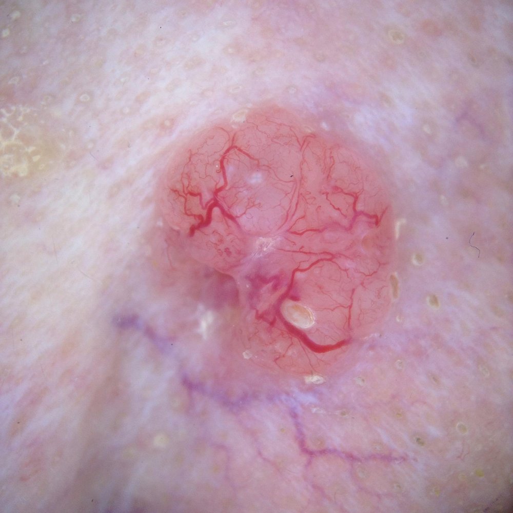 _Nodular basal cell carcinoma: a pearly, translucent pink nodule with fine branching (arborizing) blood vessels — a classic appearance._
## How it is diagnosed
Diagnosis starts with a skin exam, often using a handheld magnifier (dermoscopy). The diagnosis is confirmed with a **biopsy**, where a small sample is taken and checked under the microscope. The biopsy also tells the type, or subtype, of BCC, which guides treatment.
Tumors are then sorted into **lower-risk** and **higher-risk** groups. That group drives the choice of treatment. A BCC is higher-risk if it has an aggressive type under the microscope, sits on the central face, is large, has fuzzy borders, has come back before, or grows in someone with a weak immune system.
## How it is treated
Most basal cell carcinomas are cured with treatment done in the office. The right option depends on the tumor's risk group, size, location, and your preferences. Here is how the options compare, strongest evidence first.
**Surgery is the standard** for most BCC. It has the lowest recurrence rates of any option. For **high-risk, recurrent, or central-face tumors**, a margin-controlled technique called Mohs surgery checks the whole edge of the removed tissue under the microscope during the visit. In a 10-year trial of high-risk facial BCC, recurrence after standard surgery was about 12% for new tumors and 14% for returning ones; with the margin-controlled technique it was about 4%. The difference was clear for returning tumors but not for new ones. Guidelines save the margin-controlled approach for those higher-risk cases.
For **small, low-risk surface tumors**, other options can work when surgery is declined or not practical. These include a prescription cream (imiquimod), freezing or scraping-and-cautery, and light treatment (photodynamic therapy). They clear fewer tumors and come back more often than surgery, but can leave a better cosmetic result. **Radiation** is an option too, often for older patients or when surgery is not a good fit, though it comes back more than surgery.
A small number of BCCs grow deep or spread and cannot be treated by surgery or radiation. These are treated with pills (hedgehog inhibitors) or, in some cases, immunotherapy. That care is given through a referral to a specialist who treats systemic cancer (a medical oncologist).
## Lowering your risk and catching new ones early
Most basal cell carcinoma is caused by ultraviolet light from the sun and tanning beds, so reducing UV exposure over a lifetime lowers the risk. For people who have already had two or more skin cancers, a daily vitamin (nicotinamide) modestly lowered the rate of new non-melanoma skin cancers in a trial.
Having one basal cell carcinoma raises the chance of getting another, and some recurrences appear more than five years later, so regular skin checks are an important part of care after a diagnosis.
## When to see a doctor
See a clinician for any spot that is new, changing, bleeding, or not healing, especially a sore that heals and comes back in the same place. If you have already had a basal cell carcinoma, keep up with regular skin checks, since new spots and late recurrences can appear years later.
## Frequently asked questions
**Is basal cell carcinoma dangerous?**
It is rarely life-threatening because it almost never spreads to other organs. The concern is local growth: left untreated, it can damage nearby skin, cartilage, and tissue, especially on the face. Caught early, it is highly curable.
**Will it come back after treatment?**
Most tumors are cured, and surgery has the lowest recurrence rates. Some recurrences appear more than five years later, and having one BCC raises the chance of getting another, so ongoing skin checks matter.
**Can I treat it with a cream instead of surgery?**
For small, low-risk surface tumors, a prescription cream is an option, but it cures fewer tumors than surgery. In a head-to-head trial, 3-year success was 84% with imiquimod cream vs 98% with surgery. Creams are not used for higher-risk or deeper tumors.
**What is Mohs surgery, and do I need it?**
It is a margin-controlled technique that checks the whole edge of the removed tissue under the microscope during the procedure. Guidelines reserve it for high-risk, recurrent, or critical-site tumors rather than every BCC. Your clinician can tell you whether your tumor falls into that group.
**Does basal cell carcinoma spread?**
Very rarely. Spread to lymph nodes or other organs happens in a small fraction of cases. The usual concern is the tumor growing into nearby tissue, not spreading through the body.
## Questions to ask your clinician
- What risk group is my tumor, and how does that change my options?
- Which treatment fits this spot, and what will the scar or healed area look like?
- Will the removed tissue be checked for clear edges?
- How often should I have skin checks now that I have had a BCC?
- What can I do to lower my risk of new skin cancers?
Basal cell carcinoma is one of several skin cancers and precancers. Related guides cover [squamous cell carcinoma](/clinical-guides/squamous-cell-carcinoma), [actinic keratosis](/clinical-guides/actinic-keratosis), and the treatments above, including [surgical excision](/clinical-guides/surgical-excision). A thorough [skin cancer screening](/blog/skin-cancer-screening-the-villages) is the place to start.
_Reviewed by Tyler Long, DO. Last reviewed 2026-06-24._
### Melanoma
https://spotdoc.com/clinical-guides/melanoma
Melanoma is the **most serious common skin cancer** because it can spread to other parts of the body. But here is the hopeful part. Most melanomas are caught early, and an early melanoma is **highly treatable**. This guide is an overview of the whole melanoma family. It explains the types and stages, so you can see where a diagnosis fits, then walks through how treatment changes from one stage to the next.
## Key points
- Melanoma is a **family** of cancers, from melanoma in situ (top layer only, almost always curable) to invasive and advanced disease. Where a melanoma falls drives everything that follows.
- Most melanoma is **caught early and cured with a single surgery**. Advanced disease has powerful newer treatments, given by oncology.
- **Outlook depends most on how early it is found.** A new or changing mole is the key warning sign.
## What is melanoma?
Melanoma is a cancer of the **melanocytes**, the cells that give skin its color. It can appear in an existing mole or as a new spot. It shows up most on sun-exposed skin, but it can occur anywhere, including the soles, palms, and nails. The main risk factor is ultraviolet light, along with having many moles, fair skin, and a family history. What sets melanoma apart from other skin cancers is that it is more likely to **spread** if it is not caught early.
## The melanoma family: where a diagnosis fits
Melanoma is not one thing. The biggest difference between melanomas is **how deep the cancer has grown**, and that is what places a diagnosis on the map below:
- **Melanoma in situ (Stage 0).** The earliest form. The cancer cells sit only in the top layer of skin and have not grown deeper, so it essentially cannot spread and is almost always cured by removing it. The [melanoma in situ](/clinical-guides/melanoma-in-situ) guide covers this. A common in-situ type on chronically sun-damaged skin, often on the face in older adults, is **lentigo maligna** (see the [lentigo maligna](/clinical-guides/lentigo-maligna) guide).
- **Invasive melanoma (Stage I–II).** The melanoma has grown down into the skin but has not spread. It is staged mainly by its **depth** (the Breslow thickness). Surgery is the main treatment, and most are cured.
- **Advanced melanoma (Stage III–IV).** The melanoma has spread, either to nearby lymph nodes (Stage III) or to distant parts of the body (Stage IV). Treatment adds systemic therapy, given by oncology.
Melanoma also comes in **subtypes**, named for how it looks and grows: **superficial spreading** (the most common, spreads outward first), **nodular** (grows downward faster, so it can be thicker when found), **lentigo maligna** (on sun-damaged skin in older adults), and **acral lentiginous** (on the palms, soles, or under the nails). For treatment, the stage matters more than the subtype, but the subtype affects where to look and how it appears.
## Warning signs: the ABCDEs
A new or changing mole is the most important clue. The **ABCDE** guide helps spot a worrisome mole:
- **A**symmetry: one half does not match the other
- **B**order: edges are ragged, notched, or blurred
- **C**olor: more than one color, or uneven color
- **D**iameter: larger than a pencil eraser (about 6 mm), though melanomas can be smaller
- **E**volving: changing in size, shape, color, or feel, or a new spot
Another simple clue is the "ugly duckling": a mole that looks different from your others. Anything new, changing, bleeding, or itchy is worth getting checked.
## How it is diagnosed and staged
A clinician examines the spot, often with a handheld magnifier (dermoscopy). Any suspicious lesion is removed and checked under the microscope (a biopsy).
If it is melanoma, the most important number is the **Breslow thickness**. This is how deep the melanoma reaches into the skin, in millimeters. Thin melanomas have the best outlook. Thickness, plus whether the surface is broken (ulceration), sets the **stage**, which guides treatment. The current staging system (AJCC 8th edition) is built so that the small share of higher-stage tumors accounts for most of the serious outcomes.
## Treatment, by stage
Treatment follows the stage. Here is the short version of each; the deeper guides cover the details.
### Melanoma in situ
Early, surface-only melanoma is removed by surgery with a narrow margin, and it is almost always cured. For lentigo maligna on the face, when surgery is difficult, nonsurgical options such as a skin cream or radiation are sometimes used. The [melanoma in situ](/clinical-guides/melanoma-in-situ) guide covers this in full.
### Invasive melanoma
For invasive melanoma, **surgery is the main treatment**. The melanoma is removed with a margin of normal skin matched to how deep it is, and most are cured. (A dedicated invasive melanoma guide, with the full treatment detail, is in progress.)
Beyond surgery, a few other treatments are used in **selected cases** and are led by oncology. They are not first-line, but worth knowing about:
- **Radiation** can be added after surgery in certain higher-risk situations, used when surgery is not a good option, or given to ease symptoms.
- **Neoadjuvant immunotherapy** means immunotherapy given *before* surgery, for higher-risk melanoma that can still be removed. It is a newer approach, studied in recent trials and used in selected patients.
- **Intralesional or limb-directed therapy** uses an injection into the tumor, or treatment delivered to an affected arm or leg. It can be used when melanoma comes back in the skin near the original site ("in-transit" disease).
### Checking the lymph nodes
For thicker melanomas, a **sentinel lymph node biopsy** may be offered. This checks the first lymph node the melanoma would drain to. It is mainly a **staging** tool: it gives the strongest single clue about outlook and helps decide whether more treatment is considered. It is not a treatment that extends survival for everyone. If that node has melanoma, doctors now often watch the area with ultrasound rather than removing all the nodes, because removing them did not improve survival in a large trial.
### Advanced melanoma
For advanced melanoma (spread to nodes or beyond), treatment is systemic and given by oncology:
- **Immunotherapy** helps the immune system attack the melanoma. After surgery for higher-risk disease it lowers the chance of return, and for spread melanoma it has meaningfully improved long-term survival.
- **Targeted pills (BRAF/MEK inhibitors)** are an option for the roughly half of melanomas that carry a BRAF gene change, found by gene testing.
This care is coordinated by referral to a medical oncologist.
## Lowering your risk and catching it early
Most melanoma risk traces back to ultraviolet light, so sun protection lowers the chance of new melanoma over time. Because outlook depends so much on early detection, knowing your own skin and getting new or changing spots checked are the most powerful things you can do.
Checking your own skin regularly, and having higher-risk skin watched by a clinician, helps catch melanoma when it is thin and most treatable. See the [skin self-exam](/clinical-guides/skin-self-exam) guide for how to do it.
## When to see a doctor
See a clinician for any mole or spot that is new, changing, asymmetric, multi-colored, growing, bleeding, or simply looks different from your others. Do not wait for it to hurt. If you have many moles, atypical (dysplastic) moles, fair skin, or a family history of melanoma, regular skin checks are especially worthwhile.
## Frequently asked questions
**What is the difference between melanoma in situ and invasive melanoma?**
Melanoma in situ sits only in the top layer of skin, so it essentially cannot spread and is almost always cured by removing it. Invasive melanoma has grown deeper into the skin, which is why it is staged by depth and why it can, if not caught early, spread. They are treated differently, so it is worth knowing which one you have.
**Is melanoma curable?**
Most early melanomas are cured with a single surgery. Outlook depends most on how early it is found and how thin the melanoma is, which is why checking new or changing spots matters so much.
**Will I need chemotherapy?**
For most early melanoma, no. Treatment is a surgical removal in the office. Advanced disease is treated with immunotherapy or targeted pills, not old-style chemotherapy, and that care is given by oncology.
**What is a sentinel lymph node biopsy?**
It checks the first lymph node the melanoma would drain to. It is mainly a staging tool that gives the strongest clue about outlook and helps guide further treatment, offered for thicker melanomas. If that node is involved, watching the nodes with ultrasound is now often chosen, since removing all of them did not improve survival in a trial.
**How wide a margin will be removed?**
The margin is matched to how deep the melanoma is: thin melanomas need a narrower margin than thicker ones. Trials show a margin matched to the tumor is as safe as a wider one. Your clinician will explain the plan for your melanoma.
## Questions to ask your clinician
- Is my melanoma in situ, invasive, or advanced, and what stage is it?
- How thick is it, and do I need a sentinel lymph node biopsy?
- What margin will be removed, and what will the scar look like?
- Do I need to see an oncologist, and for what?
- How often should I have skin checks now, and what should I watch for?
Melanoma is the most serious skin cancer, but early detection changes everything. Related guides cover [melanoma in situ](/clinical-guides/melanoma-in-situ), [lentigo maligna](/clinical-guides/lentigo-maligna), [atypical (dysplastic) moles](/clinical-guides/atypical-moles), and [surgical excision](/clinical-guides/surgical-excision). A thorough [skin cancer screening](/blog/skin-cancer-screening-the-villages) is the place to start.
_Reviewed by Tyler Long, DO. Last reviewed 2026-06-24._
### Melanoma In Situ
https://spotdoc.com/clinical-guides/melanoma-in-situ
Melanoma in situ is the earliest form of melanoma — "in situ" means it's still confined to the epidermis, the top layer of skin. It has not grown into the deeper layers where it could reach blood vessels or lymphatics, so while it remains in situ it cannot spread. That's why, despite the word "melanoma," the outlook is excellent: removing it completely is essentially curative.
## Key points
- Melanoma in situ is **Stage 0** — confined to the surface, with no ability to spread while it stays in situ.
- **Removing it completely is essentially curative**, usually with a minor outpatient excision.
- It's a signal to **watch your skin closely** afterward, since one skin cancer raises the odds of another.
If you've been told you have melanoma in situ, this is the reassuring end of the melanoma spectrum — but it still deserves prompt, complete treatment and ongoing skin checks.
## What is melanoma in situ?
Melanoma begins in melanocytes, the pigment-producing cells of the skin. In melanoma in situ, those abnormal cells are still contained within the epidermis. Left alone over time, some can progress to invasive melanoma, which is why it's treated rather than watched. A closely related subtype that arises on chronically sun-damaged skin — typically the face of older adults — is [lentigo maligna](/clinical-guides/lentigo-maligna), which behaves differently enough to have its own guide.
## Signs to watch for
It usually looks like a new or changing flat spot or patch, often tan to brown to black, sometimes with uneven color or an irregular border — the **ABCDEs** (asymmetry, border, color, diameter, evolving). Many are found during a routine skin exam before they cause any symptom at all, which is the case for early detection.
## How it's diagnosed
A suspicious spot is examined, often with a dermoscope, and confirmed with a **skin biopsy** — the only way to diagnose melanoma. The pathologist confirms the cells are confined to the epidermis (in situ) rather than invasive, which is what places it at Stage 0.
## What treatment usually looks like
Because the abnormal cells sit only at the surface, the goal is simply to remove them completely. For most lesions that means **surgical excision with a small margin of normal-looking skin**, done in the office under local anesthesia — and that's typically the end of it. For lesions with hard-to-see borders, a margin-controlled (staged) approach may be used to confirm the edges are clear before closing.
Which approach your dermatologist recommends depends on the lesion's size, location, and how well-defined its borders are. There's no chemotherapy and no systemic treatment for in-situ disease — the work is local and, once margins are clear, done. Surgery is the standard, but the right plan is a shared decision with your doctor: for most people that means complete removal, while for a few — for example, someone with very limited life expectancy, or where an operation would carry more risk than a slow-growing surface lesion — careful monitoring can be a reasonable choice made together.
## Margins: why a little extra skin is removed
When the spot is removed, your doctor also takes a thin rim of normal-looking skin around it. That rim is called the **margin**. A **clear margin** means no abnormal cells are left at the edge.
Melanoma in situ can spread sideways under the skin, a little past what the eye can see. So a very small margin is not always enough. In one large study, a 5 mm margin cleared all the cells in about 8 out of 10 cases, while about 9 mm cleared nearly all of them. Your doctor checks the edges under a microscope. If any cells reach the edge, a little more skin is removed until the margin is clear. This is why complete removal works so well: once the edges are clear, the spot is gone.
## Do I need scans or a lymph node test?
No. Because the cells stay in the top layer of skin, melanoma in situ cannot reach the lymph nodes or other organs. So body scans and a lymph node biopsy are not needed. Those tests are used for melanoma that has grown deeper. If a biopsy ever shows the melanoma went past "in situ," your care would change, and your doctor would talk you through the next steps.
## Reducing your risk going forward
Having had a melanoma in situ raises your future risk, so the fundamentals matter more than ever: broad-spectrum sun protection, no tanning beds, monthly self-checks, and regular [skin checks](/blog/skin-cancer-screening-the-villages). For people with many moles, [total body photography](/clinical-guides/total-body-photography) creates a baseline to track changes over time.
## When to see a doctor
Have any new, changing, or unusual spot evaluated promptly — and keep your scheduled skin checks, since the most useful thing after one skin cancer is catching the next one early.
## Frequently asked questions
**Is melanoma in situ dangerous?**
While it stays in situ it cannot spread, and complete removal is essentially curative. It's treated promptly because, left alone, some lesions can progress to invasive melanoma over time.
**Will I need anything besides surgery?**
Usually not. In-situ disease is treated locally — there's no role for chemotherapy or systemic therapy. Ongoing skin surveillance is the main follow-up.
**How is this different from lentigo maligna?**
Lentigo maligna is a subtype of melanoma in situ on sun-damaged skin (usually the face) that spreads more widely under the surface, so it often needs wider or margin-controlled removal. See the [lentigo maligna guide](/clinical-guides/lentigo-maligna).
**Can melanoma in situ spread to other parts of my body?**
Not while it stays "in situ." The cells sit in the top layer of skin, with no path to the blood or lymph nodes. The reason to treat it now is to remove it before it ever has the chance to grow deeper.
**How big will the scar be?**
The area removed is a bit larger than the spot, because of the margin of normal skin around it. Most wounds close in a thin line. Spots on the face or larger lesions may need a more careful repair.
**Can it come back after it's removed?**
When the whole lesion is removed with clear margins, it rarely comes back, about 1 in 100. More often, a "new" spot later is a separate skin cancer, which is why ongoing skin checks matter.
**How often should I get my skin checked afterward?**
Most people have a full skin exam at least once a year for life, and sometimes more often at first. Your dermatologist sets the timing based on your skin, your moles, and your family history.
## Questions to ask your dermatologist
- Were my margins clear, or do I need more skin removed?
- Was the melanoma only in situ, or did any of it go deeper?
- How often should I have my skin checked from now on?
- What changes should make me call the office sooner?
_Reviewed by Tyler Long, DO. Last updated June 2026._
### Photodynamic Therapy (PDT)
https://spotdoc.com/clinical-guides/photodynamic-therapy
Photodynamic therapy (PDT) is an in-office treatment that uses a **light-activated medicine** to clear sun-damaged and precancerous skin. It is most often used for [actinic keratoses](/clinical-guides/actinic-keratosis), the rough, scaly precancers caused by years of sun, and it treats a whole sun-damaged area at once rather than freezing spots one at a time.
## Key points
- PDT uses a **light-activated medicine** to destroy sun-damaged and precancerous cells across a whole area.
- It is mainly used for **actinic keratoses** and usually heals with a **good cosmetic result**.
- A gentler **daylight** version clears about as well as the in-office lamp, with much less stinging.
## What is photodynamic therapy?
PDT combines two things: a medicine and light. A cream or liquid is put on the skin, the damaged cells soak it up, and then a special light switches it on to destroy those cells. Because healthy skin absorbs far less of the medicine, the effect is concentrated where the sun damage is. It is non-surgical, with no cut and no stitches.
## How it works
1. **The medicine.** A photosensitizer (a light-sensitive medicine such as aminolevulinic acid, or ALA) is applied to the skin. Rapidly dividing, sun-damaged cells take it up and turn it into a compound that makes them sensitive to light.
2. **Incubation.** The medicine is left on for a while so it builds up in the abnormal cells.
3. **Light.** The area is exposed to a specific light (often blue or red). The light activates the medicine, which produces a burst of oxygen that destroys the targeted cells.
## What PDT treats
PDT treats sun-damaged and early-cancerous skin across a whole area at once, rather than one spot at a time. It is used most for **[actinic keratoses](/clinical-guides/actinic-keratosis)**, especially when there are many in one area ("field cancerization"). Here is what it treats and how well the research shows it works, strongest first.
Clearing a sun-damaged field is also a form of skin cancer prevention, because some actinic keratoses, left alone, can turn into squamous cell carcinoma. PDT is **not** used for invasive (deeper) skin cancers or for melanoma, which need other treatments.
## What to expect at your visit
A session is straightforward but takes a couple of hours start to finish:
- **Prep.** The skin is cleaned and sometimes lightly exfoliated so the medicine absorbs evenly.
- **Medicine applied.** The photosensitizer is put on the treatment area.
- **Incubation.** You wait while it absorbs, from a short period up to a couple of hours.
- **Light.** You sit under the light for about 10 to 20 minutes. Many people feel stinging or burning during this step; a fan or cool air helps.
- **Home.** It is done in one visit, though a full course often includes a **second session** a few weeks later for the best clearance.
## Side effects and recovery
Afterward the treated skin behaves like it has a strong sunburn: **redness, swelling, stinging, then crusting and peeling**, which usually settles over about a week as fresh skin comes through. The most important aftercare rule is **strict sun avoidance for about 48 hours**, because the medicine leaves your skin very light-sensitive and ordinary daylight, even through a window, can cause a strong reaction during that window.
## Conventional vs. daylight PDT
There are two ways to deliver the light:
- **Conventional (in-office lamp) PDT** uses a blue or red light in the clinic. It is effective but can sting during the light exposure.
- **Daylight PDT** uses natural daylight to activate the medicine instead of a lamp. Studies find it clears actinic keratoses **about as well as the lamp while causing much less pain**, with the trade-off that it depends on suitable weather and a specific exposure window.
## Blue light vs. red light
The activating light comes in two colors, and which one is used depends on what is being treated:
- **Blue light** works mainly at the skin's surface. It is the common choice for **actinic keratoses** on the face and scalp.
- **Red light** reaches a little deeper into the skin, so it is often chosen for **thicker spots or early skin cancers** such as Bowen's disease and superficial basal cell carcinoma.
For actinic keratoses, the two work about equally well. In a head-to-head randomized trial, blue and red light cleared a similar share of people (about 85 percent), with only small differences in stinging.
## How well it works
The table above summarizes the evidence by condition. The detail below adds the key numbers.
For **actinic keratoses**, PDT clears most treated spots. In one large study about **89% of people had a strong response** by week 12, and a newer gel cleared spots in about **78% of people**. Head-to-head with **freezing**, PDT clears about the same share of spots (roughly 7 in 10) but leaves a **better cosmetic result** and higher satisfaction. The gentler **daylight** version clears about as well as the in-office lamp with much less pain. Major guidelines list PDT as an effective option for actinic keratoses.
For **Bowen's disease**, a trial found PDT cleared about **80% of patches at one year, more than freezing (67%) or 5-FU cream (69%)**, with the best cosmetic result of the three. For **superficial basal cell carcinoma**, PDT clears most thin lesions and looks better than surgery, but surgery has fewer recurrences, so PDT is reserved for selected, surface-level cases.
For how PDT stacks up against creams and freezing for actinic keratoses, see the [actinic keratosis guide](/clinical-guides/actinic-keratosis).
## Is PDT right for you?
PDT is a strong fit for people with **many actinic keratoses** or widespread sun damage in one area, where freezing each spot would be impractical. It is one of several field treatments; others include the creams **5-fluorouracil** and **imiquimod**, compared side by side in the [actinic keratosis guide](/clinical-guides/actinic-keratosis). The best choice depends on how much sun damage you have, your schedule, and how your skin tolerates treatment. A thorough [skin cancer screening](/blog/skin-cancer-screening-the-villages) is the place to start that conversation.
## Frequently asked questions
**Does photodynamic therapy hurt?**
The light step can sting or burn, and a fan or cool air helps. The gentler daylight version is nearly painless. Afterward the skin feels like a sunburn for a few days.
**How many sessions will I need?**
Most people have two sessions a few weeks apart for the best clearance, then return for touch-ups over time as new sun damage appears.
**How long is the downtime?**
The treated skin is red, then crusts and peels, usually settling over about a week. Plan for strict sun avoidance for about 48 hours right after.
**Will PDT get rid of all my spots?**
It clears most treated spots, but not always every one, and new spots can form later from ongoing sun damage. Ongoing sun protection and regular skin checks remain important.
**Is PDT used for skin cancer?**
It is mainly for actinic keratoses, and in selected cases for superficial basal cell carcinoma and squamous cell carcinoma in situ (Bowen's disease). It is not used for invasive skin cancers or melanoma.
## Questions to ask your dermatologist
- Is PDT a better fit for me than a field cream or freezing?
- Will I need one session or two?
- What should I expect for redness and downtime, and when can I be back in the sun?
- How will we keep an eye on the area afterward?
_Reviewed by Tyler Long, DO. Last updated June 2026._
### Skin Self-Exam (ABCDE)
https://spotdoc.com/clinical-guides/skin-self-exam
A skin self-exam is a simple, regular check of your own skin for new or changing spots. The goal is not to diagnose anything yourself. The goal is to notice a spot worth showing a doctor, so a skin cancer can be found while it is small and easy to treat. This guide explains how to check your skin with the ABCDE rule and the ugly duckling sign, what a self-exam can miss, and gives honest answers about the phone apps people ask about.
## Key points
- Knowing your own skin helps you find a new or changing spot early, when treatment is simpler.
- The two tools to use are the **ABCDE rule** and the **ugly duckling sign**, the spot that looks different from your others.
- A self-exam is **one layer** of early detection, not the whole plan. It does not replace a professional skin check, and self-detected spots are more often missed on the back.
## What a skin self-exam is
A skin self-exam means looking over your whole body every so often to learn what is normal for you, then watching for anything that is new, changing, or simply does not fit. The single most useful thing you can notice is **change**. A spot that grows, darkens, changes shape, itches, or bleeds is worth a closer look.
To check well, use good light and a full-length mirror, and use a hand mirror for your back and scalp. Look everywhere, including places the sun rarely reaches: your scalp, behind your ears, between your toes, the soles of your feet, and under your nails. Many spots are easy to see in a mirror, but the back is a common blind spot, which is one reason a partner helps.
## How to check: ABCDE and the ugly duckling
Two simple guides cover most of what to look for.
The **ABCDE rule** lists the warning signs of melanoma:
- **A is for Asymmetry.** One half does not match the other.
- **B is for Border.** The edges are uneven, notched, or blurred.
- **C is for Color.** More than one color, or an uneven mix of brown, black, red, white, or blue.
- **D is for Diameter.** Larger than a pencil eraser, about 6 millimeters, though some melanomas are smaller.
- **E is for Evolving.** Any change in size, shape, color, or feel. The letter E was added because change is such a strong warning sign.
The **ugly duckling sign** is even simpler. Most of your moles tend to look alike. The one that stands out from the rest, the odd one, is the one to show a doctor. In studies, melanomas usually stood out as the different spot when compared with a person's other moles.
## What the research shows about self-exams
Here is each part of a self-exam and how well it is supported, graded on the same scale used across this library.
A few honest notes on this table. Finding skin cancer earlier, when it is thinner, is clearly tied to better outcomes. What is not proven is that checking your own skin lowers the chance of dying from melanoma. One early study suggested a large benefit, but the authors said it needed to be repeated, and a 20-year follow-up of the same people did not confirm it. A review of the few trials that exist could neither prove nor rule out a benefit from skin checks. So a self-exam is best seen as useful skin awareness that works alongside professional exams, not as a test that stands on its own. It also misses things. Self-detected melanomas are more often thicker than those a doctor finds, especially on the back, which is hard to see.
## Other options patients ask about
People often arrive having downloaded an app or planning to keep an eye on a spot on their own. Here are the honest answers.
- **"Can a phone app check my mole for me?"** Not reliably. When researchers tested mole-check apps, the results were poor and uneven. One app missed every melanoma it was shown, and the apps as a group could not be relied on to catch all skin cancers. A review of dozens of these apps found that none had been properly tested for accuracy. An app may remind you to look, but it cannot tell you a spot is safe. If a spot worries you, see a clinician, whatever an app says.
- **"Should I track my own photos?"** Yes, as a memory aid. Comparing photos of a spot over time helps you and your doctor notice change, the strongest warning sign. Just remember a photo is a way to track a spot, not a way to diagnose it.
- **"Can I remove a worrying spot myself?"** No. At-home mole-removal pens, creams, and burning kits do nothing to find skin cancer, and they destroy the very tissue a doctor needs to tell whether a spot was harmless. Checking and removing are different things, and removal belongs in a clinic.
- **"Can I just ask an online symptom checker?"** General symptom-checker tools and chatbots are not tested to rule out skin cancer. Treat a reassuring answer as meaningless for a spot that is new, changing, or different.
## What a self-exam can miss
The ABCDE rule and the ugly duckling sign are good for the common type of melanoma, the flat, dark, uneven kind. They are weaker for other kinds, so it helps to know the gaps.
- **Fast-growing bumps.** Some melanomas are raised, firm, and one even color. A spot that is growing quickly or feels different deserves attention even if it passes the ABCDE check.
- **Skin-colored or pink spots.** Not every melanoma is dark. Some are pink, red, or skin-colored, which makes them easy to overlook.
- **Palms, soles, and nails.** Melanoma can appear on the palms, the soles of the feet, between the toes, and as a new dark streak under a nail. These spots are easy to miss, so check them on purpose.
This matters for everyone, and it matters for people with brown and black skin. Melanoma is less common in darker skin, but it is often found at a later stage. Outcome gaps for melanoma on the hands and feet are real and are not explained by late diagnosis alone. The takeaway is the same for all skin tones: check everywhere, including soles and nails, and get a new, changing, bleeding, or non-healing spot looked at, even if a rule or an app says it looks fine.
## When to see a doctor
A self-exam is a way to decide when to call, not a way to clear a spot yourself. Have a clinician check any spot that is new, growing, changing color, bleeding, itching, or not healing. Show them the spot that looks different from your others. If you have fair skin, a lot of past sun, many moles, or a personal or family history of skin cancer, ask about a regular professional skin exam. Catching a skin cancer early is still the most dependable way to keep treatment small and simple.
## Frequently asked questions
**How often should I check my skin?**
Many dermatology groups, including the American Academy of Dermatology, suggest a self-check about once a month, which is a reasonable rhythm for most people. No study has proven that a particular schedule lowers the risk of dying from skin cancer, so the real goal is to check often enough to know what is normal for you and to notice change. Pick a routine you will keep, and look more often if you are at higher risk or watching a particular spot.
**Does checking my own skin lower my risk of dying from melanoma?**
There is no randomized trial proving that. What is clear is that skin cancer found earlier, while it is thinner, is tied to better outcomes. A self-exam is useful awareness that works best alongside professional skin exams, not a replacement for them.
**What is the ugly duckling sign?**
Most of your moles look alike. The ugly duckling is the spot that stands out as different from the rest. That odd one is the one to show a doctor, even if it does not clearly break the ABCDE rule.
**Can a mole-check app tell me if a spot is cancer?**
No. When these apps were tested, the results were poor and uneven, and some missed melanomas entirely. An app may help you remember to look, but it cannot confirm a spot is safe. See a clinician for anything that worries you.
**Where do people most often miss a melanoma?**
On hard-to-see areas, especially the back, and on places people forget to check, like the scalp, soles, between the toes, and under the nails. A mirror and a partner help cover these spots.
## Questions to ask your dermatologist
- Given my skin type and history, how should I check my own skin?
- How often should I have a professional skin exam?
- Which of my spots should I watch, and what change should make me call?
- Should I take baseline photos of any spots to track them?
- What kinds of melanoma might a simple self-check miss for me?
_Reviewed by Tyler Long, DO. Last reviewed June 2026._
### Squamous Cell Carcinoma
https://spotdoc.com/clinical-guides/squamous-cell-carcinoma
Squamous cell carcinoma (SCC) is the **second most common skin cancer**. It comes from years of ultraviolet light. It often starts in a precancer called an [actinic keratosis](/clinical-guides/actinic-keratosis). Most tumors are found early and cured with surgery. But unlike basal cell carcinoma, some higher-risk tumors can **spread**, usually first to nearby lymph nodes. That is why risk features and early treatment matter.
## Key points
- SCC is the **second most common skin cancer**; it comes from sun damage and grows faster than basal cell carcinoma.
- **Most are cured with surgery.** A higher-risk minority can spread, so the tumor's risk features guide how it is treated and followed.
- A **biopsy** confirms it, and people with a weak immune system are higher-risk and need closer skin checks.
## What is squamous cell carcinoma?
SCC is a cancer of the flat (squamous) cells in the outer layer of skin. The main cause is years of ultraviolet light from the sun or tanning beds. It often grows on the most sun-exposed areas: the face, ears, lips, scalp, and backs of the hands. It is common, with hundreds of thousands of cases each year in the United States.
Most squamous cell carcinomas are cured by removing them. They are taken seriously because some higher-risk tumors can grow deep and spread, usually first to nearby lymph nodes. Spread is uncommon overall. It is more likely with certain tumors, so the risk features below shape the plan.
## Signs and what to look for
Squamous cell carcinoma can show up in several ways. Any spot that is new, growing, tender, or not healing is worth checking. Common signs include:
- A **firm red bump** or nodule
- A **rough, scaly, or crusted patch** that may bleed
- A **sore that does not heal**, or heals and comes back
- A **wart-like growth** or a tender, raised spot, sometimes on the lip or ear
A spot that grows quickly, hurts, or bleeds easily is worth getting looked at sooner.
_Nodular basal cell carcinoma: a pearly, translucent pink nodule with fine branching (arborizing) blood vessels — a classic appearance._
## How it is diagnosed
Diagnosis starts with a skin exam, often using a handheld magnifier (dermoscopy). The diagnosis is confirmed with a **biopsy**, where a small sample is taken and checked under the microscope. The biopsy also tells the type, or subtype, of BCC, which guides treatment.
Tumors are then sorted into **lower-risk** and **higher-risk** groups. That group drives the choice of treatment. A BCC is higher-risk if it has an aggressive type under the microscope, sits on the central face, is large, has fuzzy borders, has come back before, or grows in someone with a weak immune system.
## How it is treated
Most basal cell carcinomas are cured with treatment done in the office. The right option depends on the tumor's risk group, size, location, and your preferences. Here is how the options compare, strongest evidence first.
**Surgery is the standard** for most BCC. It has the lowest recurrence rates of any option. For **high-risk, recurrent, or central-face tumors**, a margin-controlled technique called Mohs surgery checks the whole edge of the removed tissue under the microscope during the visit. In a 10-year trial of high-risk facial BCC, recurrence after standard surgery was about 12% for new tumors and 14% for returning ones; with the margin-controlled technique it was about 4%. The difference was clear for returning tumors but not for new ones. Guidelines save the margin-controlled approach for those higher-risk cases.
For **small, low-risk surface tumors**, other options can work when surgery is declined or not practical. These include a prescription cream (imiquimod), freezing or scraping-and-cautery, and light treatment (photodynamic therapy). They clear fewer tumors and come back more often than surgery, but can leave a better cosmetic result. **Radiation** is an option too, often for older patients or when surgery is not a good fit, though it comes back more than surgery.
A small number of BCCs grow deep or spread and cannot be treated by surgery or radiation. These are treated with pills (hedgehog inhibitors) or, in some cases, immunotherapy. That care is given through a referral to a specialist who treats systemic cancer (a medical oncologist).
## Lowering your risk and catching new ones early
Most basal cell carcinoma is caused by ultraviolet light from the sun and tanning beds, so reducing UV exposure over a lifetime lowers the risk. For people who have already had two or more skin cancers, a daily vitamin (nicotinamide) modestly lowered the rate of new non-melanoma skin cancers in a trial.
Having one basal cell carcinoma raises the chance of getting another, and some recurrences appear more than five years later, so regular skin checks are an important part of care after a diagnosis.
## When to see a doctor
See a clinician for any spot that is new, changing, bleeding, or not healing, especially a sore that heals and comes back in the same place. If you have already had a basal cell carcinoma, keep up with regular skin checks, since new spots and late recurrences can appear years later.
## Frequently asked questions
**Is basal cell carcinoma dangerous?**
It is rarely life-threatening because it almost never spreads to other organs. The concern is local growth: left untreated, it can damage nearby skin, cartilage, and tissue, especially on the face. Caught early, it is highly curable.
**Will it come back after treatment?**
Most tumors are cured, and surgery has the lowest recurrence rates. Some recurrences appear more than five years later, and having one BCC raises the chance of getting another, so ongoing skin checks matter.
**Can I treat it with a cream instead of surgery?**
For small, low-risk surface tumors, a prescription cream is an option, but it cures fewer tumors than surgery. In a head-to-head trial, 3-year success was 84% with imiquimod cream vs 98% with surgery. Creams are not used for higher-risk or deeper tumors.
**What is Mohs surgery, and do I need it?**
It is a margin-controlled technique that checks the whole edge of the removed tissue under the microscope during the procedure. Guidelines reserve it for high-risk, recurrent, or critical-site tumors rather than every BCC. Your clinician can tell you whether your tumor falls into that group.
**Does basal cell carcinoma spread?**
Very rarely. Spread to lymph nodes or other organs happens in a small fraction of cases. The usual concern is the tumor growing into nearby tissue, not spreading through the body.
## Questions to ask your clinician
- What risk group is my tumor, and how does that change my options?
- Which treatment fits this spot, and what will the scar or healed area look like?
- Will the removed tissue be checked for clear edges?
- How often should I have skin checks now that I have had a BCC?
- What can I do to lower my risk of new skin cancers?
Basal cell carcinoma is one of several skin cancers and precancers. Related guides cover [squamous cell carcinoma](/clinical-guides/squamous-cell-carcinoma), [actinic keratosis](/clinical-guides/actinic-keratosis), and the treatments above, including [surgical excision](/clinical-guides/surgical-excision). A thorough [skin cancer screening](/blog/skin-cancer-screening-the-villages) is the place to start.
_Reviewed by Tyler Long, DO. Last reviewed 2026-06-24._
### Melanoma
https://spotdoc.com/clinical-guides/melanoma
Melanoma is the **most serious common skin cancer** because it can spread to other parts of the body. But here is the hopeful part. Most melanomas are caught early, and an early melanoma is **highly treatable**. This guide is an overview of the whole melanoma family. It explains the types and stages, so you can see where a diagnosis fits, then walks through how treatment changes from one stage to the next.
## Key points
- Melanoma is a **family** of cancers, from melanoma in situ (top layer only, almost always curable) to invasive and advanced disease. Where a melanoma falls drives everything that follows.
- Most melanoma is **caught early and cured with a single surgery**. Advanced disease has powerful newer treatments, given by oncology.
- **Outlook depends most on how early it is found.** A new or changing mole is the key warning sign.
## What is melanoma?
Melanoma is a cancer of the **melanocytes**, the cells that give skin its color. It can appear in an existing mole or as a new spot. It shows up most on sun-exposed skin, but it can occur anywhere, including the soles, palms, and nails. The main risk factor is ultraviolet light, along with having many moles, fair skin, and a family history. What sets melanoma apart from other skin cancers is that it is more likely to **spread** if it is not caught early.
## The melanoma family: where a diagnosis fits
Melanoma is not one thing. The biggest difference between melanomas is **how deep the cancer has grown**, and that is what places a diagnosis on the map below:
- **Melanoma in situ (Stage 0).** The earliest form. The cancer cells sit only in the top layer of skin and have not grown deeper, so it essentially cannot spread and is almost always cured by removing it. The [melanoma in situ](/clinical-guides/melanoma-in-situ) guide covers this. A common in-situ type on chronically sun-damaged skin, often on the face in older adults, is **lentigo maligna** (see the [lentigo maligna](/clinical-guides/lentigo-maligna) guide).
- **Invasive melanoma (Stage I–II).** The melanoma has grown down into the skin but has not spread. It is staged mainly by its **depth** (the Breslow thickness). Surgery is the main treatment, and most are cured.
- **Advanced melanoma (Stage III–IV).** The melanoma has spread, either to nearby lymph nodes (Stage III) or to distant parts of the body (Stage IV). Treatment adds systemic therapy, given by oncology.
Melanoma also comes in **subtypes**, named for how it looks and grows: **superficial spreading** (the most common, spreads outward first), **nodular** (grows downward faster, so it can be thicker when found), **lentigo maligna** (on sun-damaged skin in older adults), and **acral lentiginous** (on the palms, soles, or under the nails). For treatment, the stage matters more than the subtype, but the subtype affects where to look and how it appears.
## Warning signs: the ABCDEs
A new or changing mole is the most important clue. The **ABCDE** guide helps spot a worrisome mole:
- **A**symmetry: one half does not match the other
- **B**order: edges are ragged, notched, or blurred
- **C**olor: more than one color, or uneven color
- **D**iameter: larger than a pencil eraser (about 6 mm), though melanomas can be smaller
- **E**volving: changing in size, shape, color, or feel, or a new spot
Another simple clue is the "ugly duckling": a mole that looks different from your others. Anything new, changing, bleeding, or itchy is worth getting checked.
## How it is diagnosed and staged
A clinician examines the spot, often with a handheld magnifier (dermoscopy). Any suspicious lesion is removed and checked under the microscope (a biopsy).
If it is melanoma, the most important number is the **Breslow thickness**. This is how deep the melanoma reaches into the skin, in millimeters. Thin melanomas have the best outlook. Thickness, plus whether the surface is broken (ulceration), sets the **stage**, which guides treatment. The current staging system (AJCC 8th edition) is built so that the small share of higher-stage tumors accounts for most of the serious outcomes.
## Treatment, by stage
Treatment follows the stage. Here is the short version of each; the deeper guides cover the details.
### Melanoma in situ
Early, surface-only melanoma is removed by surgery with a narrow margin, and it is almost always cured. For lentigo maligna on the face, when surgery is difficult, nonsurgical options such as a skin cream or radiation are sometimes used. The [melanoma in situ](/clinical-guides/melanoma-in-situ) guide covers this in full.
### Invasive melanoma
For invasive melanoma, **surgery is the main treatment**. The melanoma is removed with a margin of normal skin matched to how deep it is, and most are cured. (A dedicated invasive melanoma guide, with the full treatment detail, is in progress.)
Beyond surgery, a few other treatments are used in **selected cases** and are led by oncology. They are not first-line, but worth knowing about:
- **Radiation** can be added after surgery in certain higher-risk situations, used when surgery is not a good option, or given to ease symptoms.
- **Neoadjuvant immunotherapy** means immunotherapy given *before* surgery, for higher-risk melanoma that can still be removed. It is a newer approach, studied in recent trials and used in selected patients.
- **Intralesional or limb-directed therapy** uses an injection into the tumor, or treatment delivered to an affected arm or leg. It can be used when melanoma comes back in the skin near the original site ("in-transit" disease).
### Checking the lymph nodes
For thicker melanomas, a **sentinel lymph node biopsy** may be offered. This checks the first lymph node the melanoma would drain to. It is mainly a **staging** tool: it gives the strongest single clue about outlook and helps decide whether more treatment is considered. It is not a treatment that extends survival for everyone. If that node has melanoma, doctors now often watch the area with ultrasound rather than removing all the nodes, because removing them did not improve survival in a large trial.
### Advanced melanoma
For advanced melanoma (spread to nodes or beyond), treatment is systemic and given by oncology:
- **Immunotherapy** helps the immune system attack the melanoma. After surgery for higher-risk disease it lowers the chance of return, and for spread melanoma it has meaningfully improved long-term survival.
- **Targeted pills (BRAF/MEK inhibitors)** are an option for the roughly half of melanomas that carry a BRAF gene change, found by gene testing.
This care is coordinated by referral to a medical oncologist.
## Lowering your risk and catching it early
Most melanoma risk traces back to ultraviolet light, so sun protection lowers the chance of new melanoma over time. Because outlook depends so much on early detection, knowing your own skin and getting new or changing spots checked are the most powerful things you can do.
Checking your own skin regularly, and having higher-risk skin watched by a clinician, helps catch melanoma when it is thin and most treatable. See the [skin self-exam](/clinical-guides/skin-self-exam) guide for how to do it.
## When to see a doctor
See a clinician for any mole or spot that is new, changing, asymmetric, multi-colored, growing, bleeding, or simply looks different from your others. Do not wait for it to hurt. If you have many moles, atypical (dysplastic) moles, fair skin, or a family history of melanoma, regular skin checks are especially worthwhile.
## Frequently asked questions
**What is the difference between melanoma in situ and invasive melanoma?**
Melanoma in situ sits only in the top layer of skin, so it essentially cannot spread and is almost always cured by removing it. Invasive melanoma has grown deeper into the skin, which is why it is staged by depth and why it can, if not caught early, spread. They are treated differently, so it is worth knowing which one you have.
**Is melanoma curable?**
Most early melanomas are cured with a single surgery. Outlook depends most on how early it is found and how thin the melanoma is, which is why checking new or changing spots matters so much.
**Will I need chemotherapy?**
For most early melanoma, no. Treatment is a surgical removal in the office. Advanced disease is treated with immunotherapy or targeted pills, not old-style chemotherapy, and that care is given by oncology.
**What is a sentinel lymph node biopsy?**
It checks the first lymph node the melanoma would drain to. It is mainly a staging tool that gives the strongest clue about outlook and helps guide further treatment, offered for thicker melanomas. If that node is involved, watching the nodes with ultrasound is now often chosen, since removing all of them did not improve survival in a trial.
**How wide a margin will be removed?**
The margin is matched to how deep the melanoma is: thin melanomas need a narrower margin than thicker ones. Trials show a margin matched to the tumor is as safe as a wider one. Your clinician will explain the plan for your melanoma.
## Questions to ask your clinician
- Is my melanoma in situ, invasive, or advanced, and what stage is it?
- How thick is it, and do I need a sentinel lymph node biopsy?
- What margin will be removed, and what will the scar look like?
- Do I need to see an oncologist, and for what?
- How often should I have skin checks now, and what should I watch for?
Melanoma is the most serious skin cancer, but early detection changes everything. Related guides cover [melanoma in situ](/clinical-guides/melanoma-in-situ), [lentigo maligna](/clinical-guides/lentigo-maligna), [atypical (dysplastic) moles](/clinical-guides/atypical-moles), and [surgical excision](/clinical-guides/surgical-excision). A thorough [skin cancer screening](/blog/skin-cancer-screening-the-villages) is the place to start.
_Reviewed by Tyler Long, DO. Last reviewed 2026-06-24._
### Melanoma In Situ
https://spotdoc.com/clinical-guides/melanoma-in-situ
Melanoma in situ is the earliest form of melanoma — "in situ" means it's still confined to the epidermis, the top layer of skin. It has not grown into the deeper layers where it could reach blood vessels or lymphatics, so while it remains in situ it cannot spread. That's why, despite the word "melanoma," the outlook is excellent: removing it completely is essentially curative.
## Key points
- Melanoma in situ is **Stage 0** — confined to the surface, with no ability to spread while it stays in situ.
- **Removing it completely is essentially curative**, usually with a minor outpatient excision.
- It's a signal to **watch your skin closely** afterward, since one skin cancer raises the odds of another.
If you've been told you have melanoma in situ, this is the reassuring end of the melanoma spectrum — but it still deserves prompt, complete treatment and ongoing skin checks.
## What is melanoma in situ?
Melanoma begins in melanocytes, the pigment-producing cells of the skin. In melanoma in situ, those abnormal cells are still contained within the epidermis. Left alone over time, some can progress to invasive melanoma, which is why it's treated rather than watched. A closely related subtype that arises on chronically sun-damaged skin — typically the face of older adults — is [lentigo maligna](/clinical-guides/lentigo-maligna), which behaves differently enough to have its own guide.
## Signs to watch for
It usually looks like a new or changing flat spot or patch, often tan to brown to black, sometimes with uneven color or an irregular border — the **ABCDEs** (asymmetry, border, color, diameter, evolving). Many are found during a routine skin exam before they cause any symptom at all, which is the case for early detection.
## How it's diagnosed
A suspicious spot is examined, often with a dermoscope, and confirmed with a **skin biopsy** — the only way to diagnose melanoma. The pathologist confirms the cells are confined to the epidermis (in situ) rather than invasive, which is what places it at Stage 0.
## What treatment usually looks like
Because the abnormal cells sit only at the surface, the goal is simply to remove them completely. For most lesions that means **surgical excision with a small margin of normal-looking skin**, done in the office under local anesthesia — and that's typically the end of it. For lesions with hard-to-see borders, a margin-controlled (staged) approach may be used to confirm the edges are clear before closing.
Which approach your dermatologist recommends depends on the lesion's size, location, and how well-defined its borders are. There's no chemotherapy and no systemic treatment for in-situ disease — the work is local and, once margins are clear, done. Surgery is the standard, but the right plan is a shared decision with your doctor: for most people that means complete removal, while for a few — for example, someone with very limited life expectancy, or where an operation would carry more risk than a slow-growing surface lesion — careful monitoring can be a reasonable choice made together.
## Margins: why a little extra skin is removed
When the spot is removed, your doctor also takes a thin rim of normal-looking skin around it. That rim is called the **margin**. A **clear margin** means no abnormal cells are left at the edge.
Melanoma in situ can spread sideways under the skin, a little past what the eye can see. So a very small margin is not always enough. In one large study, a 5 mm margin cleared all the cells in about 8 out of 10 cases, while about 9 mm cleared nearly all of them. Your doctor checks the edges under a microscope. If any cells reach the edge, a little more skin is removed until the margin is clear. This is why complete removal works so well: once the edges are clear, the spot is gone.
## Do I need scans or a lymph node test?
No. Because the cells stay in the top layer of skin, melanoma in situ cannot reach the lymph nodes or other organs. So body scans and a lymph node biopsy are not needed. Those tests are used for melanoma that has grown deeper. If a biopsy ever shows the melanoma went past "in situ," your care would change, and your doctor would talk you through the next steps.
## Reducing your risk going forward
Having had a melanoma in situ raises your future risk, so the fundamentals matter more than ever: broad-spectrum sun protection, no tanning beds, monthly self-checks, and regular [skin checks](/blog/skin-cancer-screening-the-villages). For people with many moles, [total body photography](/clinical-guides/total-body-photography) creates a baseline to track changes over time.
## When to see a doctor
Have any new, changing, or unusual spot evaluated promptly — and keep your scheduled skin checks, since the most useful thing after one skin cancer is catching the next one early.
## Frequently asked questions
**Is melanoma in situ dangerous?**
While it stays in situ it cannot spread, and complete removal is essentially curative. It's treated promptly because, left alone, some lesions can progress to invasive melanoma over time.
**Will I need anything besides surgery?**
Usually not. In-situ disease is treated locally — there's no role for chemotherapy or systemic therapy. Ongoing skin surveillance is the main follow-up.
**How is this different from lentigo maligna?**
Lentigo maligna is a subtype of melanoma in situ on sun-damaged skin (usually the face) that spreads more widely under the surface, so it often needs wider or margin-controlled removal. See the [lentigo maligna guide](/clinical-guides/lentigo-maligna).
**Can melanoma in situ spread to other parts of my body?**
Not while it stays "in situ." The cells sit in the top layer of skin, with no path to the blood or lymph nodes. The reason to treat it now is to remove it before it ever has the chance to grow deeper.
**How big will the scar be?**
The area removed is a bit larger than the spot, because of the margin of normal skin around it. Most wounds close in a thin line. Spots on the face or larger lesions may need a more careful repair.
**Can it come back after it's removed?**
When the whole lesion is removed with clear margins, it rarely comes back, about 1 in 100. More often, a "new" spot later is a separate skin cancer, which is why ongoing skin checks matter.
**How often should I get my skin checked afterward?**
Most people have a full skin exam at least once a year for life, and sometimes more often at first. Your dermatologist sets the timing based on your skin, your moles, and your family history.
## Questions to ask your dermatologist
- Were my margins clear, or do I need more skin removed?
- Was the melanoma only in situ, or did any of it go deeper?
- How often should I have my skin checked from now on?
- What changes should make me call the office sooner?
_Reviewed by Tyler Long, DO. Last updated June 2026._
### Photodynamic Therapy (PDT)
https://spotdoc.com/clinical-guides/photodynamic-therapy
Photodynamic therapy (PDT) is an in-office treatment that uses a **light-activated medicine** to clear sun-damaged and precancerous skin. It is most often used for [actinic keratoses](/clinical-guides/actinic-keratosis), the rough, scaly precancers caused by years of sun, and it treats a whole sun-damaged area at once rather than freezing spots one at a time.
## Key points
- PDT uses a **light-activated medicine** to destroy sun-damaged and precancerous cells across a whole area.
- It is mainly used for **actinic keratoses** and usually heals with a **good cosmetic result**.
- A gentler **daylight** version clears about as well as the in-office lamp, with much less stinging.
## What is photodynamic therapy?
PDT combines two things: a medicine and light. A cream or liquid is put on the skin, the damaged cells soak it up, and then a special light switches it on to destroy those cells. Because healthy skin absorbs far less of the medicine, the effect is concentrated where the sun damage is. It is non-surgical, with no cut and no stitches.
## How it works
1. **The medicine.** A photosensitizer (a light-sensitive medicine such as aminolevulinic acid, or ALA) is applied to the skin. Rapidly dividing, sun-damaged cells take it up and turn it into a compound that makes them sensitive to light.
2. **Incubation.** The medicine is left on for a while so it builds up in the abnormal cells.
3. **Light.** The area is exposed to a specific light (often blue or red). The light activates the medicine, which produces a burst of oxygen that destroys the targeted cells.
## What PDT treats
PDT treats sun-damaged and early-cancerous skin across a whole area at once, rather than one spot at a time. It is used most for **[actinic keratoses](/clinical-guides/actinic-keratosis)**, especially when there are many in one area ("field cancerization"). Here is what it treats and how well the research shows it works, strongest first.
Clearing a sun-damaged field is also a form of skin cancer prevention, because some actinic keratoses, left alone, can turn into squamous cell carcinoma. PDT is **not** used for invasive (deeper) skin cancers or for melanoma, which need other treatments.
## What to expect at your visit
A session is straightforward but takes a couple of hours start to finish:
- **Prep.** The skin is cleaned and sometimes lightly exfoliated so the medicine absorbs evenly.
- **Medicine applied.** The photosensitizer is put on the treatment area.
- **Incubation.** You wait while it absorbs, from a short period up to a couple of hours.
- **Light.** You sit under the light for about 10 to 20 minutes. Many people feel stinging or burning during this step; a fan or cool air helps.
- **Home.** It is done in one visit, though a full course often includes a **second session** a few weeks later for the best clearance.
## Side effects and recovery
Afterward the treated skin behaves like it has a strong sunburn: **redness, swelling, stinging, then crusting and peeling**, which usually settles over about a week as fresh skin comes through. The most important aftercare rule is **strict sun avoidance for about 48 hours**, because the medicine leaves your skin very light-sensitive and ordinary daylight, even through a window, can cause a strong reaction during that window.
## Conventional vs. daylight PDT
There are two ways to deliver the light:
- **Conventional (in-office lamp) PDT** uses a blue or red light in the clinic. It is effective but can sting during the light exposure.
- **Daylight PDT** uses natural daylight to activate the medicine instead of a lamp. Studies find it clears actinic keratoses **about as well as the lamp while causing much less pain**, with the trade-off that it depends on suitable weather and a specific exposure window.
## Blue light vs. red light
The activating light comes in two colors, and which one is used depends on what is being treated:
- **Blue light** works mainly at the skin's surface. It is the common choice for **actinic keratoses** on the face and scalp.
- **Red light** reaches a little deeper into the skin, so it is often chosen for **thicker spots or early skin cancers** such as Bowen's disease and superficial basal cell carcinoma.
For actinic keratoses, the two work about equally well. In a head-to-head randomized trial, blue and red light cleared a similar share of people (about 85 percent), with only small differences in stinging.
## How well it works
The table above summarizes the evidence by condition. The detail below adds the key numbers.
For **actinic keratoses**, PDT clears most treated spots. In one large study about **89% of people had a strong response** by week 12, and a newer gel cleared spots in about **78% of people**. Head-to-head with **freezing**, PDT clears about the same share of spots (roughly 7 in 10) but leaves a **better cosmetic result** and higher satisfaction. The gentler **daylight** version clears about as well as the in-office lamp with much less pain. Major guidelines list PDT as an effective option for actinic keratoses.
For **Bowen's disease**, a trial found PDT cleared about **80% of patches at one year, more than freezing (67%) or 5-FU cream (69%)**, with the best cosmetic result of the three. For **superficial basal cell carcinoma**, PDT clears most thin lesions and looks better than surgery, but surgery has fewer recurrences, so PDT is reserved for selected, surface-level cases.
For how PDT stacks up against creams and freezing for actinic keratoses, see the [actinic keratosis guide](/clinical-guides/actinic-keratosis).
## Is PDT right for you?
PDT is a strong fit for people with **many actinic keratoses** or widespread sun damage in one area, where freezing each spot would be impractical. It is one of several field treatments; others include the creams **5-fluorouracil** and **imiquimod**, compared side by side in the [actinic keratosis guide](/clinical-guides/actinic-keratosis). The best choice depends on how much sun damage you have, your schedule, and how your skin tolerates treatment. A thorough [skin cancer screening](/blog/skin-cancer-screening-the-villages) is the place to start that conversation.
## Frequently asked questions
**Does photodynamic therapy hurt?**
The light step can sting or burn, and a fan or cool air helps. The gentler daylight version is nearly painless. Afterward the skin feels like a sunburn for a few days.
**How many sessions will I need?**
Most people have two sessions a few weeks apart for the best clearance, then return for touch-ups over time as new sun damage appears.
**How long is the downtime?**
The treated skin is red, then crusts and peels, usually settling over about a week. Plan for strict sun avoidance for about 48 hours right after.
**Will PDT get rid of all my spots?**
It clears most treated spots, but not always every one, and new spots can form later from ongoing sun damage. Ongoing sun protection and regular skin checks remain important.
**Is PDT used for skin cancer?**
It is mainly for actinic keratoses, and in selected cases for superficial basal cell carcinoma and squamous cell carcinoma in situ (Bowen's disease). It is not used for invasive skin cancers or melanoma.
## Questions to ask your dermatologist
- Is PDT a better fit for me than a field cream or freezing?
- Will I need one session or two?
- What should I expect for redness and downtime, and when can I be back in the sun?
- How will we keep an eye on the area afterward?
_Reviewed by Tyler Long, DO. Last updated June 2026._
### Skin Self-Exam (ABCDE)
https://spotdoc.com/clinical-guides/skin-self-exam
A skin self-exam is a simple, regular check of your own skin for new or changing spots. The goal is not to diagnose anything yourself. The goal is to notice a spot worth showing a doctor, so a skin cancer can be found while it is small and easy to treat. This guide explains how to check your skin with the ABCDE rule and the ugly duckling sign, what a self-exam can miss, and gives honest answers about the phone apps people ask about.
## Key points
- Knowing your own skin helps you find a new or changing spot early, when treatment is simpler.
- The two tools to use are the **ABCDE rule** and the **ugly duckling sign**, the spot that looks different from your others.
- A self-exam is **one layer** of early detection, not the whole plan. It does not replace a professional skin check, and self-detected spots are more often missed on the back.
## What a skin self-exam is
A skin self-exam means looking over your whole body every so often to learn what is normal for you, then watching for anything that is new, changing, or simply does not fit. The single most useful thing you can notice is **change**. A spot that grows, darkens, changes shape, itches, or bleeds is worth a closer look.
To check well, use good light and a full-length mirror, and use a hand mirror for your back and scalp. Look everywhere, including places the sun rarely reaches: your scalp, behind your ears, between your toes, the soles of your feet, and under your nails. Many spots are easy to see in a mirror, but the back is a common blind spot, which is one reason a partner helps.
## How to check: ABCDE and the ugly duckling
Two simple guides cover most of what to look for.
The **ABCDE rule** lists the warning signs of melanoma:
- **A is for Asymmetry.** One half does not match the other.
- **B is for Border.** The edges are uneven, notched, or blurred.
- **C is for Color.** More than one color, or an uneven mix of brown, black, red, white, or blue.
- **D is for Diameter.** Larger than a pencil eraser, about 6 millimeters, though some melanomas are smaller.
- **E is for Evolving.** Any change in size, shape, color, or feel. The letter E was added because change is such a strong warning sign.
The **ugly duckling sign** is even simpler. Most of your moles tend to look alike. The one that stands out from the rest, the odd one, is the one to show a doctor. In studies, melanomas usually stood out as the different spot when compared with a person's other moles.
## What the research shows about self-exams
Here is each part of a self-exam and how well it is supported, graded on the same scale used across this library.
A few honest notes on this table. Finding skin cancer earlier, when it is thinner, is clearly tied to better outcomes. What is not proven is that checking your own skin lowers the chance of dying from melanoma. One early study suggested a large benefit, but the authors said it needed to be repeated, and a 20-year follow-up of the same people did not confirm it. A review of the few trials that exist could neither prove nor rule out a benefit from skin checks. So a self-exam is best seen as useful skin awareness that works alongside professional exams, not as a test that stands on its own. It also misses things. Self-detected melanomas are more often thicker than those a doctor finds, especially on the back, which is hard to see.
## Other options patients ask about
People often arrive having downloaded an app or planning to keep an eye on a spot on their own. Here are the honest answers.
- **"Can a phone app check my mole for me?"** Not reliably. When researchers tested mole-check apps, the results were poor and uneven. One app missed every melanoma it was shown, and the apps as a group could not be relied on to catch all skin cancers. A review of dozens of these apps found that none had been properly tested for accuracy. An app may remind you to look, but it cannot tell you a spot is safe. If a spot worries you, see a clinician, whatever an app says.
- **"Should I track my own photos?"** Yes, as a memory aid. Comparing photos of a spot over time helps you and your doctor notice change, the strongest warning sign. Just remember a photo is a way to track a spot, not a way to diagnose it.
- **"Can I remove a worrying spot myself?"** No. At-home mole-removal pens, creams, and burning kits do nothing to find skin cancer, and they destroy the very tissue a doctor needs to tell whether a spot was harmless. Checking and removing are different things, and removal belongs in a clinic.
- **"Can I just ask an online symptom checker?"** General symptom-checker tools and chatbots are not tested to rule out skin cancer. Treat a reassuring answer as meaningless for a spot that is new, changing, or different.
## What a self-exam can miss
The ABCDE rule and the ugly duckling sign are good for the common type of melanoma, the flat, dark, uneven kind. They are weaker for other kinds, so it helps to know the gaps.
- **Fast-growing bumps.** Some melanomas are raised, firm, and one even color. A spot that is growing quickly or feels different deserves attention even if it passes the ABCDE check.
- **Skin-colored or pink spots.** Not every melanoma is dark. Some are pink, red, or skin-colored, which makes them easy to overlook.
- **Palms, soles, and nails.** Melanoma can appear on the palms, the soles of the feet, between the toes, and as a new dark streak under a nail. These spots are easy to miss, so check them on purpose.
This matters for everyone, and it matters for people with brown and black skin. Melanoma is less common in darker skin, but it is often found at a later stage. Outcome gaps for melanoma on the hands and feet are real and are not explained by late diagnosis alone. The takeaway is the same for all skin tones: check everywhere, including soles and nails, and get a new, changing, bleeding, or non-healing spot looked at, even if a rule or an app says it looks fine.
## When to see a doctor
A self-exam is a way to decide when to call, not a way to clear a spot yourself. Have a clinician check any spot that is new, growing, changing color, bleeding, itching, or not healing. Show them the spot that looks different from your others. If you have fair skin, a lot of past sun, many moles, or a personal or family history of skin cancer, ask about a regular professional skin exam. Catching a skin cancer early is still the most dependable way to keep treatment small and simple.
## Frequently asked questions
**How often should I check my skin?**
Many dermatology groups, including the American Academy of Dermatology, suggest a self-check about once a month, which is a reasonable rhythm for most people. No study has proven that a particular schedule lowers the risk of dying from skin cancer, so the real goal is to check often enough to know what is normal for you and to notice change. Pick a routine you will keep, and look more often if you are at higher risk or watching a particular spot.
**Does checking my own skin lower my risk of dying from melanoma?**
There is no randomized trial proving that. What is clear is that skin cancer found earlier, while it is thinner, is tied to better outcomes. A self-exam is useful awareness that works best alongside professional skin exams, not a replacement for them.
**What is the ugly duckling sign?**
Most of your moles look alike. The ugly duckling is the spot that stands out as different from the rest. That odd one is the one to show a doctor, even if it does not clearly break the ABCDE rule.
**Can a mole-check app tell me if a spot is cancer?**
No. When these apps were tested, the results were poor and uneven, and some missed melanomas entirely. An app may help you remember to look, but it cannot confirm a spot is safe. See a clinician for anything that worries you.
**Where do people most often miss a melanoma?**
On hard-to-see areas, especially the back, and on places people forget to check, like the scalp, soles, between the toes, and under the nails. A mirror and a partner help cover these spots.
## Questions to ask your dermatologist
- Given my skin type and history, how should I check my own skin?
- How often should I have a professional skin exam?
- Which of my spots should I watch, and what change should make me call?
- Should I take baseline photos of any spots to track them?
- What kinds of melanoma might a simple self-check miss for me?
_Reviewed by Tyler Long, DO. Last reviewed June 2026._
### Squamous Cell Carcinoma
https://spotdoc.com/clinical-guides/squamous-cell-carcinoma
Squamous cell carcinoma (SCC) is the **second most common skin cancer**. It comes from years of ultraviolet light. It often starts in a precancer called an [actinic keratosis](/clinical-guides/actinic-keratosis). Most tumors are found early and cured with surgery. But unlike basal cell carcinoma, some higher-risk tumors can **spread**, usually first to nearby lymph nodes. That is why risk features and early treatment matter.
## Key points
- SCC is the **second most common skin cancer**; it comes from sun damage and grows faster than basal cell carcinoma.
- **Most are cured with surgery.** A higher-risk minority can spread, so the tumor's risk features guide how it is treated and followed.
- A **biopsy** confirms it, and people with a weak immune system are higher-risk and need closer skin checks.
## What is squamous cell carcinoma?
SCC is a cancer of the flat (squamous) cells in the outer layer of skin. The main cause is years of ultraviolet light from the sun or tanning beds. It often grows on the most sun-exposed areas: the face, ears, lips, scalp, and backs of the hands. It is common, with hundreds of thousands of cases each year in the United States.
Most squamous cell carcinomas are cured by removing them. They are taken seriously because some higher-risk tumors can grow deep and spread, usually first to nearby lymph nodes. Spread is uncommon overall. It is more likely with certain tumors, so the risk features below shape the plan.
## Signs and what to look for
Squamous cell carcinoma can show up in several ways. Any spot that is new, growing, tender, or not healing is worth checking. Common signs include:
- A **firm red bump** or nodule
- A **rough, scaly, or crusted patch** that may bleed
- A **sore that does not heal**, or heals and comes back
- A **wart-like growth** or a tender, raised spot, sometimes on the lip or ear
A spot that grows quickly, hurts, or bleeds easily is worth getting looked at sooner.
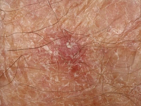 _Squamous cell carcinoma: a firm, red, scaly patch with crusting on sun-damaged skin, a common appearance on the face, ears, scalp, or backs of the hands._
## How it is diagnosed
Diagnosis starts with a skin exam and is confirmed with a **biopsy**, where a small sample is checked under the microscope. The biopsy shows the type and grade of the tumor, which helps sort it into a risk group.
Some tumors are **higher-risk**, which means they are more likely to come back or spread. A tumor is higher-risk if it is large, grows deep, is a higher grade, grows along a nerve, sits on the ear or lip, or grows in someone with a weak immune system. Higher-risk tumors may need imaging or a check of the lymph nodes, plus closer follow-up.
## How it is treated
Most squamous cell carcinomas are cured with treatment done in the office. The choice depends on the tumor's risk group, size, and location. Here is how the options compare, strongest evidence first.
**Surgery is the standard** first-line treatment for most localized tumors. A small margin of normal skin clears most tumors, with a wider margin for high-risk ones. For **high-risk, recurrent, or critical-site tumors**, a margin-controlled technique, known as Mohs surgery, checks the entire edge of the removed tissue under the microscope during the procedure; guidelines reserve it for those situations.
**Radiation** is an option when surgery is not possible, and it is sometimes added after surgery when a margin cannot be cleared or a tumor has grown along a nerve. Scraping-and-cautery or freezing can treat selected small, low-risk tumors, but are not used for high-risk ones.
The **rare tumor that spreads** or cannot be cured by surgery and radiation is treated with immunotherapy, given by oncology through a referral.
## Special note for people with a weakened immune system
Organ-transplant recipients and others on long-term immune-suppressing medicine get many more squamous cell carcinomas, and the tumors tend to behave more aggressively. If this is you, regular skin checks and prompt treatment of new spots are especially important, and care is usually coordinated with your transplant or specialty team.
## Lowering your risk and catching new ones early
Most of the risk comes from ultraviolet light, so sun protection lowers the chance of new skin cancers over time. Treating precancers (actinic keratoses) also helps. For high-risk patients, a daily vitamin (nicotinamide) lowered new skin cancers in a trial, and selected transplant patients may be offered a prescription retinoid.
Having one squamous cell carcinoma raises the chance of getting another, so regular skin checks are an important part of care after a diagnosis.
## When to see a doctor
See a clinician for any spot that is growing, scaly, tender, bleeding, or not healing, especially on sun-exposed skin like the face, ears, lips, scalp, or hands. If you have had a squamous cell carcinoma before, or you take immune-suppressing medicine, keep up with regular skin checks.
## Frequently asked questions
**Is squamous cell carcinoma dangerous?**
Most are cured when treated early. The concern is the higher-risk minority that can grow deep or spread, usually first to nearby lymph nodes. The tumor's risk features guide how closely it is treated and followed.
**How is it different from basal cell carcinoma?**
Both come from sun damage, but squamous cell carcinoma grows faster and is more likely to spread, while basal cell carcinoma almost never spreads. Both are usually cured with surgery when caught early.
**Will it come back?**
Most tumors are cured. Cancer left at the edge raises the chance of recurrence, so margins are checked and an involved edge is re-removed. Having one SCC also raises the chance of getting another, so ongoing skin checks matter.
**Do I need Mohs surgery?**
Not for every tumor. Mohs is a margin-controlled technique that guidelines reserve for high-risk, recurrent, or critical-site tumors. Your clinician can tell you whether your tumor falls into that group.
**Why does my immune system matter?**
People on immune-suppressing medicine, such as transplant recipients, get many more squamous cell carcinomas and more aggressive ones. They need closer skin checks and prompt treatment.
## Questions to ask your clinician
- What risk group is my tumor, and does it need imaging or a lymph node check?
- Which treatment fits this spot, and will the edges be checked for clear margins?
- Do I need radiation after surgery for any reason?
- How often should I have skin checks now?
- I take immune-suppressing medicine, so how does that change my plan?
Squamous cell carcinoma is one of several skin cancers and precancers. Related guides cover [actinic keratosis](/clinical-guides/actinic-keratosis) (its common precancer), [basal cell carcinoma](/clinical-guides/basal-cell-carcinoma), and the treatments above, including [surgical excision](/clinical-guides/surgical-excision). A thorough [skin cancer screening](/blog/skin-cancer-screening-the-villages) is the place to start.
_Reviewed by Tyler Long, DO. Last reviewed 2026-06-24._
### Sunscreen & SPF
https://spotdoc.com/clinical-guides/sunscreen
Sunscreen is a cream, lotion, or spray that blocks or absorbs the sun's ultraviolet (UV) rays before they damage your skin. Used every day, it is one of the simplest ways to lower your risk of skin cancer and to slow the wrinkling and spotting that come from sun. This guide explains what the research shows sunscreen actually prevents, how to pick and use one, and gives honest answers to the common worries about safety.
## Key points
- Daily sunscreen has been shown in a randomized trial to lower **squamous cell skin cancer**, and the benefit lasted for years.
- It also lowers **precancers** (actinic keratoses) and slows **skin aging**.
- Sunscreen is **one layer** of sun protection, not the whole plan. Shade, clothing, and avoiding midday sun matter too.
## What sunscreen is and how SPF works
Sunscreens use two kinds of filters. **Mineral** filters (zinc oxide and titanium dioxide) sit on the skin and block UV. **Chemical** filters soak up UV and turn it into a tiny amount of heat. Both kinds lower the UV that reaches your skin.
Two words on the label matter most:
- **SPF** (sun protection factor) measures protection against UVB, the rays that cause sunburn. Higher numbers block more.
- **Broad spectrum** means the product also guards against UVA, the rays tied to aging and skin cancer.
Most adults should use a broad-spectrum sunscreen of **SPF 30 or higher**. One catch: people almost always apply less than the amount used in lab tests, so the real protection is often well below the number on the bottle. In testing, typical use gave only about 20% to 50% of the labeled SPF. The fix is simple. Apply a generous layer, and reapply about every two hours and after swimming or sweating.
## What the research shows it prevents
Sunscreen is studied more than almost any prevention step in skin care. The strongest evidence comes from a long randomized trial in Australia and from large follow-up studies. Here is what regular daily use does, outcome by outcome, including where the benefit is clear and where it is not.
A few honest notes on this table. The squamous cell and precancer results are the most solid. The melanoma result leans positive but is not settled: the one randomized trial just missed the usual line for proof, and the studies that follow large groups of people are mixed.
The one result that surprises people is basal cell carcinoma, the most common but least dangerous skin cancer. The trials did not show a clear drop in it. That is not because sunscreen fails here. The reason is the *kind* of sun that drives each cancer. Squamous cell cancer and precancers track steady, lifelong sun, so cutting recent sun lowers them fairly quickly. Basal cell cancer is tied more to intense, on-and-off sun and to sunburns earlier in life, and it can take decades to appear. Starting daily sunscreen as an adult, over a few years, acts too late and too briefly to undo that early risk. Even so, basal cell rates did drift downward with longer follow-up, just not enough to be certain. So the proof is strongest for squamous cell cancer and precancers, and sunscreen is still worth using to lower basal cell risk over a lifetime, especially when started early. Sunscreen helps, but it is not a force field, which is why it works best alongside shade and clothing.
## Other options and questions patients ask about
People often arrive having read that sunscreen is harmful, or that a pill or a natural oil can replace it. Here are the honest answers.
- **"Is sunscreen toxic, or does it cause cancer?"** Studies show that some chemical filters are absorbed into the blood. That sounds alarming, but absorption is not the same as harm. The trials measured the blood levels. The researchers still said plainly that people should **not** stop using sunscreen. It means more study is needed, not that harm was found. If you would rather avoid the chemical filters, the mineral options (zinc oxide and titanium dioxide) are a simple choice. These are the two filters US regulators have proposed are safe and effective.
- **"Will sunscreen leave me low on vitamin D?"** In real life, daily sunscreen has only a small effect on vitamin D, and healthy people are unlikely to become deficient from it. A 2025 trial of strong SPF 50+ use did find a small drop in vitamin D levels and recommended a supplement for routine users. The takeaway: keep using sunscreen, and if you are concerned, get your level checked or take a vitamin D supplement.
- **"Can a pill protect me instead?"** A supplement called *Polypodium leucotomos* can modestly raise the amount of sun your skin tolerates before burning. But these studies measured sunburn thresholds, not skin cancer, and the effect is small. A pill is a possible add-on, never a replacement for sunscreen. Separately, for people who keep getting skin cancers, the vitamin **nicotinamide** (vitamin B3) has lowered new skin cancers in a trial; that is a different tool for high-risk patients, not a sunscreen substitute.
- **"Are natural oils like coconut oil a real sunscreen?"** No. Household oils do not give reliable, broad-spectrum protection, and trusting them in strong sun can leave you burned. Use a tested product with an SPF on the label.
## Does sunscreen cause melanoma? The honest answer
You may have seen the claim that sunscreen users get *more* melanoma. Some older studies did show that. It is worth understanding why. People who burn easily, sunbathe, or use tanning beds are the most likely to reach for sunscreen. They are also at higher risk of melanoma to begin with. So the sunscreen looked like the cause when the real driver was the sun-seeking. As products improved and studies got better, that false link faded. It was gone by the early 1990s. The one study type that removes this bias is the randomized trial, and it points toward benefit. The bottom line: sunscreen does not cause melanoma. But it is one layer of protection, not a reason to spend longer in the sun.
## Using sunscreen as part of a bigger plan
Sunscreen works best with the other proven steps. National guidance recommends counseling young people with fair skin on sun protection, and the same habits help adults. Build the plan around:
- **Sunscreen:** broad spectrum, SPF 30 or higher, applied well and reapplied every two hours outdoors.
- **Cover up:** a wide-brimmed hat, long sleeves, and UV-blocking sunglasses.
- **Time it:** seek shade and avoid the midday sun, about 10 a.m. to 4 p.m., and skip tanning beds.
- **Check your skin:** see a dermatologist for any new, changing, or bleeding spot, and have regular skin exams if you are at higher risk.
## When to see a doctor
Sunscreen lowers risk, but it does not erase it. Have a dermatologist check any spot that is new, growing, changing color, bleeding, or not healing. If you have had a lot of sun, fair skin, or a family history of skin cancer, ask about a yearly skin exam. Catching a skin cancer early is still the most reliable way to keep it small and easy to treat.
## Frequently asked questions
**What SPF should I use?**
Most adults should use a broad-spectrum sunscreen of SPF 30 or higher. Because people apply less than the tested amount, real protection is usually lower than the label, so apply a generous layer and reapply often.
**Is mineral or chemical sunscreen better?**
Both lower UV exposure. Mineral filters (zinc oxide, titanium dioxide) sit on the skin and are a good choice if you prefer to avoid the chemical filters. The best sunscreen is the one you will actually wear every day.
**Does sunscreen really prevent skin cancer?**
For squamous cell carcinoma and precancers, yes, that is shown in randomized trials. For melanoma the evidence leans positive but is not settled. For basal cell carcinoma the trials did not show a clear benefit, likely because that cancer is driven more by intense, on-and-off sun earlier in life and takes decades to appear. Across the board, sunscreen plus shade and clothing lowers risk.
**Will daily sunscreen make me vitamin D deficient?**
Real-life use has only a small effect, and healthy people are unlikely to become deficient. If you are worried, a vitamin D supplement or a blood test settles it, and you can keep using sunscreen.
**Do I need sunscreen on cloudy days or indoors near windows?**
UVA passes through clouds and window glass, so daily use on sun-exposed skin is reasonable if you spend time near bright windows or outdoors, even when it is overcast.
**Can a pill replace sunscreen?**
No. Oral supplements may slightly raise how much sun your skin tolerates, but they are measured against sunburn, not skin cancer, and the effect is small. Use them only as an add-on, if at all.
## Questions to ask your dermatologist
- Given my skin type and history, how much sun protection do I need?
- Is a mineral or chemical sunscreen a better fit for me?
- Should I be checked for vitamin D, or take a supplement?
- How often should I have a full skin exam?
- What kind of spot should make me call the office sooner?
_Reviewed by Tyler Long, DO. Last updated June 2026._
### Total Body Photography
https://spotdoc.com/clinical-guides/total-body-photography
Total body photography, often shortened to TBP, is a way to photograph almost all of your skin in one session and keep it as a map. At a later visit, a dermatologist can compare new pictures against that map to see what is new and what has changed. This page explains what the technology is, how it grew from flat photos to a 3D map, where the research is strongest, and what it cannot do. The close-up analysis of those images is a separate step, and it has its own page.
## Key points
- Total body photography is an imaging step. It makes a whole-body map so a new spot or a change stands out later.
- It helps most for people at higher risk of melanoma, where a new or changing spot is the main warning sign.
- Photography is the first step, not the whole exam. A dermatologist still examines your skin and takes a biopsy when a spot looks suspicious.
## What total body photography is
The idea is simple. Melanoma is often easiest to catch not from how one mole looks on a single day, but from how a spot changes over time, or from a new spot that does not match your other moles. A photographic map gives the dermatologist a fixed reference instead of relying on memory.
A modern system captures the skin from many angles at once and builds a 3D model you can rotate on a screen. Each mole sits at a fixed place on the model, so it can be found again and measured at the next visit. The images are stored as your baseline.
[Image: A 3D total body photography system, which uses an array of cameras around the patient to capture the skin surface in one session.]
## From film to a 3D map
The tools have changed a lot in twenty years. The reason to use them has not.
For a long time, tracking moles meant a doctor's memory and a few printed photos. In the 2000s this became a system. Dermatologists in Barcelona paired whole-body 2D photos with close-up pictures of single moles, an approach they called the two-step method. Over years of watching high-risk patients, they found melanomas that were mostly early. About half were the earliest, top-layer type, and the ones that had gone deeper had a median thickness near half a millimeter. A 2021 review of this kind of surveillance counted fourteen studies, and all of them used flat, 2D photos.
The same group noticed something their own numbers made hard to ignore. When they looked back at the melanomas they had caught, roughly four in ten were not moles they had been tracking. They were new spots that had appeared since the last visit. That is the gap a whole-body baseline is built to close.
3D total body photography answers that gap. Instead of a set of flat views, one session builds a full model of the skin, so today's skin can be compared against an earlier map and a new or changed spot stands out.
## How a 3D scan works, and the devices that do it
A session is quick and non-invasive. There are no injections and no radiation. It is photography. You stand in a few set poses while an array of cameras captures the skin surface in seconds. Software then stitches the images into a model, and single moles can be tagged and paired with close-up pictures for finer detail.
It helps to know that most of the published research on 3D total body photography was done on one system, the Canfield Vectra WB360. The high-risk studies from Barcelona, and the studies in Switzerland that compared 2D and 3D imaging, used it as well. Naming this matters, because it shapes what the evidence can and cannot tell us. Other tools exist. Flat 2D systems and remote mole-mapping services are still in use, and there is an academic research system from Australia, but the body of 3D evidence sits mostly with one platform.
## Where the evidence is strongest
The whole-body map earns its place in the exact spot the older flat method struggled, which is new spots.
In a 2025 study of 1,274 high-risk patients in Barcelona, almost half of the melanomas were found from the 3D map itself, before anyone used a close-up lens. An earlier study using the two-step method found that half of its melanomas were caught because of a new spot on the map or a visible change in an old one. In a large Australian high-risk clinic, new melanomas were most often first spotted with the help of the photographs.
For a person with many moles, that is the difference between hunting for one changed spot among hundreds and having the change pointed out on a picture. The melanomas found this way also tend to be thin, which is when melanoma is most treatable.
## What it does not do
The honest part matters more than the enthusiasm.
A 2025 randomized study added 3D photography and remote review to usual care and did not find a clear change in two-year results. It caught a similar number of cancers, removed fewer melanomas but more harmless spots, and cost more per person. A careful 2025 review put it plainly, that there is not yet enough evidence that 3D photography is reliable on its own. The camera also does not see everything. Studies using the 3D system note it cannot capture the scalp, the soles of the feet, or skin under clothing, so a dermatologist's eyes and hands are still needed.
Photography is the first step. What a dermatologist does with the images afterward, the close-up look at single spots and the software that can help flag a mole for a second look, is a separate step with its own page and its own limits.
## Who it is for
Total body photography is a surveillance tool, so it helps most where catching a new or changing spot early matters most. That usually means people at higher risk of [melanoma](/clinical-guides/melanoma), such as those with many moles or [atypical moles](/clinical-guides/atypical-moles), a personal or family history of melanoma, or a prior melanoma. For someone with only a few typical moles, a regular skin exam and [checking your own skin](/clinical-guides/skin-self-exam) are usually enough, and the map adds little.
## When to see a dermatologist
A baseline map does not replace paying attention between visits. See a dermatologist if you notice a spot that is new, that is changing in size, shape, or color, that looks different from your other spots, or that itches or bleeds. If you are unsure whether photography fits your risk, that is a good question to bring to a skin exam.
## Frequently asked questions
**Is total body photography the same as a skin cancer screening?**
No. It is one tool used within a skin exam, not the exam itself. A dermatologist still looks at your skin in person, examines single spots closely, and takes a biopsy when a spot is suspicious. The photographs give them a fixed reference to compare against over time.
**Does it use radiation?**
No. It is photography. There are no injections and no radiation. You stand in a few poses while cameras capture the skin surface in seconds.
**Is 3D better than the older flat photos?**
3D builds a whole-body map in one session and makes a new or changed spot easier to find. The research so far shows it helps catch new spots in high-risk patients, but a 2025 randomized study did not show a clear change in two-year results over usual care. It is a helpful tool, not a guarantee.
**Can the software find melanoma on its own?**
Not reliably yet. Software can help flag a spot for a closer look, but reviews of the evidence say it is not dependable on its own, and a dermatologist's judgment is still needed.
**Does it photograph every inch of skin?**
Almost, but not all. The scalp, the soles of the feet, and skin under clothing are not fully captured, so those areas still need a hands-on exam.
**Who benefits most?**
People at higher risk of melanoma, especially those with many moles or a personal or family history. For someone with few moles and average risk, a routine exam and self-checks usually cover it.
## Questions to ask your dermatologist
- Does my risk of melanoma make a baseline map worth it?
- How often would you repeat the imaging?
- How does the map fit with my regular skin exams and my own checks at home?
- What happens if a spot changes on the map?
## Research & news
### Can a Vitamin Lower Your Skin Cancer Risk? What the Nicotinamide Research Shows
https://spotdoc.com/blog/nicotinamide-skin-cancer
Most of what protects your skin from cancer is familiar: sunscreen, shade, and regular skin checks. So it caught attention when a large 2025 study pointed to something far simpler — an inexpensive, over-the-counter form of vitamin B3 called **nicotinamide**.
The headlines were enthusiastic, and the underlying science is genuinely encouraging. But the details matter, because nicotinamide helps some people more than others — and it is not a replacement for the basics. Here's what the research shows.
## What the 2025 Study Found
Researchers reviewed records from the U.S. Veterans Affairs health system — **33,822 patients** with a history of skin cancer — and compared those who took nicotinamide to those who didn't. The study was published in *JAMA Dermatology*.[^1]
The findings, in plain terms:
- Across everyone studied, nicotinamide was associated with about a **14% reduction** in new skin cancers.
- The effect was much larger with **timing**: patients who started nicotinamide **after their first skin cancer** saw roughly a **54% reduction** in new ones.
- The benefit was seen in **non-melanoma skin cancers** — basal cell carcinoma and squamous cell carcinoma — the keratinocyte cancers driven by sun damage.
The takeaway the authors emphasized is that **starting early matters**. The protective signal was strongest when nicotinamide began after a first skin cancer, and it weakened when started later in a long history of multiple cancers.[^2]
## This Builds on Earlier Evidence
This isn't a brand-new idea. A landmark randomized trial published in the *New England Journal of Medicine* in 2015 (the ONTRAC study) found that 500 mg of nicotinamide twice daily reduced new non-melanoma skin cancers by about 23% over a year in high-risk patients.[^3] On the strength of that trial, many dermatologists have recommended nicotinamide to selected patients for the better part of a decade. The 2025 study adds large, real-world weight to that practice.
## How It Works
Nicotinamide supports the skin in two ways that are relevant to cancer prevention. It helps replenish cellular energy (ATP) that DNA-repair machinery needs after ultraviolet damage, and it counteracts the immune suppression that UV light causes in the skin. In other words, it helps the skin repair sun damage and keep its local defenses working.
One important point of confusion: **nicotinamide is not the same as niacin.** Both are forms of vitamin B3, but niacin causes flushing and can affect cholesterol and blood pressure. Nicotinamide (also labeled niacinamide) does not cause flushing and is the form studied for skin cancer prevention. If you go looking for it, the label matters.
## Who Is Most Likely to Benefit
Based on the research, nicotinamide is most relevant for people at elevated risk of non-melanoma skin cancer — particularly those who have **already had** a [basal cell carcinoma](/clinical-guides/basal-cell-carcinoma), a [squamous cell carcinoma](/clinical-guides/squamous-cell-carcinoma), or numerous [actinic keratoses](/clinical-guides/actinic-keratosis). The dose used in the studies was **500 mg twice daily** — a standard, inexpensive over-the-counter supplement.
It is **not** established as a melanoma-prevention strategy, and it is not a reason to ease up on the fundamentals.
## What Nicotinamide Is *Not*
A few honest caveats keep this in perspective:
- **It does not replace sun protection or skin checks.** The benefit is modest-to-moderate and additive — it works alongside sunscreen, protective clothing, and regular exams, not instead of them.[^4]
- **The strongest evidence is for non-melanoma skin cancers**, not melanoma.
- **"Over-the-counter" does not mean "no conversation."** Supplements can interact with medications and conditions (for example, kidney disease can affect dosing). Talk with your dermatologist or physician before starting, and make sure you're buying nicotinamide/niacinamide — not niacin or "no-flush niacin" (inositol hexanicotinate), which are different.
- **It's a risk reducer, not a guarantee.** Even on nicotinamide, new skin cancers can still develop, which is exactly why ongoing surveillance matters.
## The Bottom Line
For the right patient — someone with a history of basal cell or squamous cell carcinoma, or extensive sun damage — nicotinamide is a low-cost, low-risk option with real evidence behind it, and the 2025 data suggest the earlier it's started, the better. It is a useful addition to a prevention plan, not a substitute for one.
If you've had a skin cancer or a lot of actinic keratoses and you're curious whether nicotinamide makes sense for you, it's a good question to raise at your next [skin cancer screening](/blog/skin-cancer-screening-the-villages).
[^1]: Breglio KF, Knox KM, Hwang J, et al. Nicotinamide for Skin Cancer Chemoprevention. *JAMA Dermatology*. 2025. [jamanetwork.com](https://jamanetwork.com/journals/jamadermatology/article-abstract/2838591)
[^2]: Vanderbilt University Medical Center. Study reveals efficacy of nicotinamide for skin cancer prevention. 2025. [news.vumc.org](https://news.vumc.org/2025/09/17/study-reveals-efficacy-of-nicotinamide-for-skin-cancer-prevention/)
[^3]: Chen AC, Martin AJ, Choy B, et al. A Phase 3 Randomized Trial of Nicotinamide for Skin-Cancer Chemoprevention. *New England Journal of Medicine*. 2015;373(17):1618–1626. [nejm.org](https://www.nejm.org/doi/full/10.1056/NEJMoa1506197)
[^4]: American Academy of Dermatology Association. Skin cancer prevention. [aad.org](https://www.aad.org/public/diseases/skin-cancer)
### Skin Cancer in the 21st Century: Takeaways From Our VHA Talk
https://spotdoc.com/blog/dermatology-21st-century-vha-lecture
This June, Dr. Tyler Long, DO, had the privilege of speaking to a full room of about 250 Villages residents at a community event hosted by the Villages Homeowners Advocates (VHA). The talk — *Dermatology of the 21st Century* — covered how skin cancer is found and treated today, and why early detection matters so much for people who live where we do.
If you couldn't make it, here are the takeaways worth keeping.
[Image: A packed house at the "Dermatology of the 21st Century" talk in The Villages — about 250 residents attended.]
## Why skin cancer hits this community harder
Skin cancer is the most common cancer in the United States — about one in five Americans will develop it in their lifetime, and roughly 9,500 people are diagnosed every day.[^1] Those numbers climb for a community like ours: older, active, and spending the day outdoors in some of the highest UV in the country.
The damage that drives skin cancer accumulates over a lifetime. A childhood up north and decades of Florida sun add up — which is exactly why the people most at risk are often the ones enjoying retirement outdoors. (We wrote more about that here: [Why Villages Residents Face a Higher Skin Cancer Risk](/blog/skin-cancer-risk-the-villages-florida).)
## The three skin cancers — and why early detection changes everything
Most skin cancer falls into three types, and all three are far easier to treat when found early:
- **Basal cell carcinoma** — the most common, slow-growing, and rarely life-threatening, but it can do real local damage if ignored.
- **Squamous cell carcinoma** — also common and highly treatable early, with more potential to spread if left alone.
- **Melanoma** — less common but the most serious. The encouraging part: when melanoma is caught at its earliest, localized stage, the five-year survival rate is about 99%.[^2]
That last figure is the whole point of the talk. Early skin cancer is usually a quick, in-office fix. The cost of waiting is what makes it dangerous.
## What to look for: the ABCDEs and the full-body skin check
A simple way to watch your own moles is the **ABCDEs**:
- **A**symmetry — one half doesn't match the other
- **B**order — edges that are ragged or blurred
- **C**olor — more than one shade, or uneven color
- **D**iameter — larger than a pencil eraser
- **E**volving — changing in size, shape, or color over time
_The ABCDEs — the warning signs to watch for in any mole._
The single most useful habit is a regular **full-body skin self-exam** — and an annual professional skin check, which looks at the places you can't easily see yourself.
## The surveillance gap: why your memory isn't enough
Here's the problem the talk spent the most time on. Skin cancer detection depends on noticing *change* — but no one, patient or physician, can reliably remember what dozens of spots looked like a year ago. That's the **surveillance gap**.
_Detection depends on noticing change over time — something memory can’t do across dozens of moles. Total body photography tracks every spot for you._
This is where [**total body photography**](/clinical-guides/total-body-photography) comes in. At SpotDoc we use a Canfield Vectra WB360 — the only one within 280 miles — to capture **92 photographs in about a second**, building a detailed map of your skin. At your next visit, we compare against that baseline so we're evaluating what's actually *changing* over time, instead of relying on memory. It's the difference between a snapshot and a track record.
_How total body photography works: capture, map, and compare over time._
## Prevention that actually works
The most effective prevention is also the least complicated:
- Daily broad-spectrum **SPF 30 or higher**, reapplied outdoors
- Sun-protective clothing, a wide-brim hat, and shade during peak hours
- No tanning beds — ever
- An annual professional skin exam
One newer point from the talk: for people with a history of certain skin cancers, **nicotinamide** — a form of vitamin B3 — has been shown in research to lower the rate of new skin cancers and now appears in national treatment guidelines. It isn't right for everyone, so ask your dermatologist whether it makes sense for you.
## Couldn't make it? Bring your questions to us
[Image: Dr. Tyler Long, DO talks with a Villages resident after the lecture.]
Some of the best moments of the evening came during the Q&A. If you have a question we didn't answer — or a spot you've been meaning to have looked at — that's exactly what we're here for. If you're overdue for a baseline skin check, you can book a scan online or call us at **(352) 914-3451**.
Thank you to the VHA and to everyone who came out. We'll see you at the next one.
[^1]: Skin Cancer Foundation — Skin Cancer Facts & Statistics. [skincancer.org](https://www.skincancer.org/skin-cancer-information/skin-cancer-facts/)
[^2]: American Cancer Society — Survival Rates for Melanoma Skin Cancer. [cancer.org](https://www.cancer.org/cancer/types/melanoma-skin-cancer/detection-diagnosis-staging/survival-rates-for-melanoma-skin-cancer-by-stage.html)
### What a New Study Reveals About Skin Cancer in Older Floridians
https://spotdoc.com/blog/skin-cancer-florida-older-adults
Most of my patients moved to Florida for the weather. The sunshine is the draw, and I get it. It's also the reason I see so much skin cancer in older adults here — and this year a study from Florida Atlantic University put hard numbers to what those of us in clinic already see.
Researchers at FAU's Charles E. Schmidt College of Medicine looked at years of national data on melanoma in Floridians aged 65 and older. The short version: it isn't getting better, and most of what's driving it can be prevented. Here's what they found, and why it matters if you live in Central Florida.
## What the Study Looked At
The team used CDC WONDER, a federal public-health database, to track new melanoma diagnoses from 2018 to 2021 and deaths from 2018 to 2023, all in Floridians 65 and up. They left out the common basal cell and squamous cell cancers on purpose, so the picture would reflect the dangerous end of the spectrum. The work was published in the *Journal of Geriatric Oncology* in May.[^1]
One number for context: Florida reports the **second-highest count of melanoma cases of any state**, behind only California.[^2] That's mostly a function of how many older, sun-exposed people live here.
## The Burden Is Holding Steady
The clearest signal in the data is how little it moves. Year over year, melanoma diagnoses and deaths in older Floridians stayed flat. No real improvement.
There was a dip in 2020. The authors chalk that up to the pandemic, when skin checks and screenings ground to a halt — fewer cancers got caught that year, even though just as many were occurring. Once screening resumed, the numbers snapped back to where they'd been.
## Older Men Are Dying About Twice as Often as Women
The finding that jumped out at me is the sex gap. In every year studied, older men died of melanoma at **roughly twice the rate of older women**.
Why? A few things stack up. Men this age are less likely to use sun protection, less likely to check their own skin, and tend to have more lifetime sun behind them; there may also be real biological differences in how the immune system handles the disease. The cause matters less than the takeaway: if you're an older man, a regular skin check earns its keep.
## Who the Data Points To
Non-Hispanic white residents had the highest rates of both diagnosis and death, which tracks with what we know about fair skin and cumulative sun. That doesn't let anyone off the hook. Melanoma can show up in any skin tone, and in darker skin it's often caught later. But it does tell us where the heaviest burden sits here.
## What the Researchers Concluded
Here's the part I appreciated. The authors didn't treat this as the unavoidable price of living somewhere sunny. They put it on persistent gaps in prevention and early detection, and called for the unglamorous stuff: routine skin checks, more awareness, real outreach to older adults.
That's good news, honestly, because every piece of it is something you can act on. Sun protection still helps even after decades outdoors: daily sunscreen, a hat, some shade. A monthly self-exam using the [ABCDE rule](/clinical-guides/melanoma) catches changes between visits. And a yearly professional exam finds what you can't — your scalp, your back, the spots you never see.
Melanoma caught before it spreads has a survival rate above 99%. After it reaches distant organs, that number drops hard.[^3] Almost all of that gap is timing, which is why early detection is worth the nuisance of an appointment.
## What This Means If You Live in The Villages
This is my patient population almost exactly: older, retired to Florida, carrying a lifetime of sun. The study is a reminder that the risk here is real and steady, and that it answers to attention.
If your last full-body skin check was more than a year ago, that's the one thing I'd move to the top of your list. Our clinic at 3614 Kiessel Road in Brownwood Square does comprehensive screening, [full-body imaging](/clinical-guides/total-body-photography), biopsies when they're needed, and ongoing care if you've had skin cancer before. We take Medicare and most major insurance, and you don't need a referral. Not sure what a visit involves? Here's [what to expect at a skin cancer screening](/blog/skin-cancer-screening-the-villages).
---
*Tyler Long, DO is a board-certified dermatologist and the lead physician at SpotDoc in The Villages. For appointments, call (352) 914-3451 or book online.*
[^1]: Srivastav M, Etzel M, Decker M, Kamm C, Lewis K, Miron E, Reis V, Sacca L. Trends in skin cancer in the Sunshine State: An ongoing concern for older adults in the United States. *Journal of Geriatric Oncology*. 2026 May;17(5):103005. [doi.org](https://doi.org/10.1016/j.jgo.2026.103005)
[^2]: Centers for Disease Control and Prevention. Skin Cancer Statistics. [cdc.gov](https://www.cdc.gov/cancer/skin/statistics/index.htm)
[^3]: American Cancer Society. Survival Rates for Melanoma Skin Cancer. [cancer.org](https://www.cancer.org/cancer/types/melanoma-skin-cancer/detection-diagnosis-staging/survival-rates-for-melanoma-skin-cancer-by-stage.html)
### Why Villages Residents Face a Higher Skin Cancer Risk
https://spotdoc.com/blog/skin-cancer-risk-the-villages-florida
Nobody moves to The Villages for the winters up north. You came for the sun, and you get it nearly every day of the year.
I spend my working days looking at what that sun does to skin. Skin cancer is more common in Central Florida than almost anywhere else in the country, and one in five Americans will develop it at some point.[^1] Around here I treat that number as a floor.
The part worth holding onto is this: skin cancer is one of the most manageable risks in medicine. It shows up on the surface of the body, where you and I can both see it. Found early, it is almost always treatable. The whole game is finding it early.
## You brought your skin history with you
Most of my patients came from the Northeast or the Midwest. Shorter summers, milder sun. All those years still count, because UV damage to skin cells adds up over a lifetime, and every bad sunburn from a Michigan childhood is still on the books. Then you retire to a state where the summer UV index regularly hits 10 or 11, and you trade an office for a golf course.
Moving to Florida in your 60s speeds that clock up considerably.
The CDC ranks Florida among the top states for melanoma incidence,[^2] and Sumter County skews older and more sun-exposed than the national average on both counts. Snowbirds get no exemption. Six months here can carry more UV than a full year did back home.
## Three cancers worth knowing by name
[Basal cell carcinoma](/clinical-guides/basal-cell-carcinoma) is the most common skin cancer, and the most common cancer of any kind. It favors the skin that gets the most sun, like the face, the ears, the scalp, and the backs of the hands. It grows slowly and rarely spreads, but ignored for years it can do real local damage. Caught early, treatment is usually simple.
[Squamous cell carcinoma](/clinical-guides/squamous-cell-carcinoma) comes second. It also favors sun-exposed skin, and unlike basal cell it can occasionally spread if ignored.
[Melanoma](/clinical-guides/melanoma) is the serious one. It can appear anywhere, including skin that never sees the sun, and it can spread quickly. When melanoma is found while it is still localized, five-year survival is over 99%. Once it has spread to distant organs, that figure falls to around 35%.[^3] The distance between those two numbers is mostly a matter of timing.
## You don't have to give up the golf
I never tell patients to stay inside. Nobody lives here for the indoors, and hiding from the sun is not required. UV exposure is the one risk factor you can control, and most of the control comes from adjusting the edges of your routine.
Timing does the heavy lifting. The sun does most of its damage between about 10 a.m. and 4 p.m., so an early tee time or an evening pickleball match takes a real bite out of your exposure, and it beats the heat besides.
Cover what you can. A wide-brimmed hat protects the ears and the back of the neck, which a visor does not. The tops of the ears are one of the most common places I find skin cancer in men. A lightweight UPF long-sleeve shirt on the course protects better than sunscreen that went on once at 8 a.m. Sunscreen still matters, to be clear: broad-spectrum, SPF 30 or higher, reapplied every two hours while you're out. Most people apply too little of it, once.
The small exposures count too. The cart ride to dinner, the dog walk, an hour on the pool deck. It all adds up over a decade, and it favors the driver's side. I see the evidence of left-side sun on faces and forearms all the time.
## Ten minutes a month
Between visits, you are the first line of detection. Once a month, after a shower, look yourself over. In a mole, you are looking for asymmetry, where one half doesn't match the other; a border that looks ragged or blurred; color that varies within the same spot; a diameter bigger than about 6mm, roughly a pencil eraser, though melanomas can be smaller; and evolution, meaning any change in size, shape, or color, or a spot that has started to bleed or itch. Dermatologists compress all of that into the mnemonic ABCDE.
Have your spouse check your back and your scalp. You can't see them, and they can. Photos help too. A phone picture of a spot today turns "has this changed?" into an easy question three months from now. We keep a full guide to the [monthly skin self-exam](/clinical-guides/skin-self-exam) if you want the complete walkthrough.
One more thing. If a spot is new, changing, bleeding, or refusing to heal, call us. Don't save it for your annual.
## Who should be extra careful
Some people start with a higher baseline.[^4] Fair skin, light eyes, or red or blond hair all mean less natural protection from UV. So does a history of sunburns, especially the blistering kind early in life, and so does a personal or family history of skin cancer. Patients with many moles, or with [atypical (dysplastic) moles](/clinical-guides/atypical-moles), need closer watching. The same goes for anyone with a suppressed immune system, whether from medication or a transplant, and for people who spent years working outdoors. Farmers, builders, veterans: this town has plenty of each.
If two or more of those describe you and your address says Florida, a yearly exam stops being optional in my book.
## What a screening here actually involves
Plan on about 45 minutes at our clinic in Brownwood Square. The visit is a head-to-toe exam. Scalp, behind the ears, between the toes, the soles of the feet. We also do [total body photography](/clinical-guides/total-body-photography) at the same visit: a ring of 92 cameras photographs your entire skin surface in about a second. That becomes a baseline map of every spot you have, and at future visits we compare against it. A new or changing spot stands out on the map even when memory swears nothing is different.
If something needs attention, we handle it in the same office. Biopsies and excisions happen here, and so does ongoing surveillance for patients who have had skin cancer before. You will not spend the next month collecting referrals across town.
We accept Medicare and most major insurance, and a screening generally doesn't require a referral. Here's [what to expect at a skin cancer screening](/blog/skin-cancer-screening-the-villages), start to finish.
## If you're new here, or here half the year
A few practical notes I give every new patient.
Get a baseline during your first season, while everything is boring. The photography is most valuable when it starts before anything is wrong.
If you're a snowbird, book your annual for a month you are reliably in town, and keep it in the same month every year. Treat it like the car service.
If you saw a dermatologist up north, ask that office to send your records, especially any biopsy reports. Knowing the spot on your shoulder was biopsied benign in 2019 saves us both time, and it may save you a needle.
That's the whole program. One exam a year, ten minutes a month, a decent hat. The sun can stay the best part of living here.
---
*Tyler Long, DO is a board-certified dermatologist and the lead physician at SpotDoc in The Villages. For appointments, call (352) 914-3451 or book online.*
[^1]: American Academy of Dermatology Association. Skin Cancer Statistics. [aad.org](https://www.aad.org/media/stats-skin-cancer)
[^2]: Centers for Disease Control and Prevention. Skin Cancer Statistics. [cdc.gov](https://www.cdc.gov/cancer/skin/statistics/index.htm)
[^3]: American Cancer Society. Survival Rates for Melanoma Skin Cancer. [cancer.org](https://www.cancer.org/cancer/types/melanoma-skin-cancer/detection-diagnosis-staging/survival-rates-for-melanoma-skin-cancer-by-stage.html)
[^4]: American Academy of Dermatology Association. Skin cancer: Risk factors. [aad.org](https://www.aad.org/public/diseases/skin-cancer)
### Skin Cancer Screening in The Villages, Florida
https://spotdoc.com/blog/skin-cancer-screening-the-villages
If you live in The Villages, you spend more time in the sun than almost anyone in the country: on the golf course, at the pool, on the pickleball court, riding the cart to dinner. That sunshine is part of what makes life here so good. It is also why a yearly skin cancer screening is one of the most worthwhile 45 minutes you can give yourself.
Skin cancer is the most common cancer in the United States, and Central Florida sees more of it than most places. The reassuring part: when it is caught early, it is almost always highly treatable.[^1] A screening is simply how you stay ahead of it. Don't settle for a flashlight exam. Here is what a thorough skin cancer screening in The Villages should look like, and how to schedule one.
## What Is a Skin Cancer Screening?
A skin cancer screening, also called a full-body skin exam or a full-body scan for skin cancer, is a visual head-to-toe examination of your skin by a trained clinician looking for spots that could be cancerous or precancerous. It is painless, takes about 45 minutes, and requires no preparation beyond removing makeup and nail polish so those areas can be seen.
A complete exam means exactly that: scalp, behind the ears, the face and neck, the back, between the fingers and toes, the soles of the feet, and other areas you cannot easily check yourself. Using a dermatoscope, a handheld magnifier with polarized light, the clinician can see pigment patterns beneath the surface that are invisible to the naked eye, which helps tell a harmless mole from one that deserves a closer look.
Most of what we find at a screening is completely benign. The point of doing it every year is to catch the rare spot that matters while it is small and simple to treat.
## Why Screening Matters Here
Skin cancer risk comes down to lifetime ultraviolet exposure, and Central Florida delivers a lot of it. The UV index here regularly reaches 10 or 11. Many Villages residents also moved from northern states, arriving with decades of accumulated sun damage before they ever saw a Florida summer. Add an active, outdoor retirement and the exposure compounds quickly. (We cover the local risk picture in more depth in [why Villages residents face a higher skin cancer risk](/blog/skin-cancer-risk-the-villages-florida).)
The three most common skin cancers behave differently, and early detection helps with all of them:
- [**Basal cell carcinoma**](/clinical-guides/basal-cell-carcinoma) is the most common. It grows slowly and rarely spreads, but left alone it can cause real local damage. Caught early, treatment is straightforward.
- [**Squamous cell carcinoma**](/clinical-guides/squamous-cell-carcinoma) is the second most common and also favors sun-exposed skin. Early detection keeps treatment simple.
- [**Melanoma**](/clinical-guides/melanoma) is less common but the most serious, because it can spread if it is not caught early. Found at an early, localized stage, the five-year survival rate is over 99%.[^2]
That last number is the whole argument for screening in one statistic. The difference between an early melanoma and a late one is largely a matter of timing, and a screening is how you control the timing.
To learn the warning signs to watch for between visits, see our guides to [melanoma](/clinical-guides/melanoma), [atypical moles](/clinical-guides/atypical-moles), and [actinic keratosis](/clinical-guides/actinic-keratosis).
## Who Should Have a Skin Cancer Screening?
For adults living in Central Florida, an annual screening is reasonable for almost everyone. It becomes especially important if any of the following apply to you:
- You have **fair skin, light eyes, or light hair**, or burn easily
- You have a **history of significant sun exposure** or blistering sunburns
- You have a **personal or family history** of melanoma or other skin cancer
- You have **many moles, or atypical (dysplastic) moles**
- You have a **weakened immune system** from medication or a medical condition
- You have noticed a **new, changing, itching, or bleeding spot**
If several of these describe you, screening is not just a good idea once in a while. It is worth keeping on an annual schedule. And if you have noticed a spot that concerns you, you do not need to wait for an annual visit; book sooner.
## What Makes a Screening at SpotDoc Different
SpotDoc is *The Skin Cancer Company*, a physician-owned practice in The Villages where skin cancer detection and treatment is the entire focus, not one service among many. That focus shows up in how we screen.
### Total Body Photography
In addition to the hands-on exam, we offer [Total Body Photography](/clinical-guides/total-body-photography) using the Canfield Vectra WB360, the only system of its kind within 280 miles. An array of cameras captures your skin surface in seconds and builds a detailed baseline map of your moles. At future visits we compare against that baseline, so a spot that is new or changing stands out objectively instead of relying on memory. For patients with many moles or a history of skin cancer, that record makes change far easier to catch.
### A board-certified dermatologist who does this all day
Your screening is performed by Tyler Long, DO, a board-certified dermatologist, alongside Kim Nguyen, NP-C. Because skin cancer is what we do, you get an exam from clinicians who look at this every day, and if something needs attention, evaluation and treatment happen in the same office.
### Detection and treatment under one roof
If a screening turns up something suspicious, we can perform a biopsy on the spot and, when treatment is needed, handle it here, including excisions, cryotherapy, electrodesiccation and curettage, and field therapies such as photodynamic therapy. You are not handed a referral and sent across town to start over.
## What Happens If We Find Something
Finding a spot at a screening is not cause for alarm. It is the system working. Here is the typical path, so there are no surprises:
1. **We take a closer look.** Using dermoscopy, we examine the spot in detail. Many lesions that look concerning at arm's length are clearly benign under magnification, and no further action is needed.
2. **We biopsy if warranted.** If a spot cannot be confidently cleared, we numb the area and take a small sample. It is a quick, minor procedure done the same day.
3. **The sample is analyzed.** A pathologist examines the tissue, and we review the results with you and explain what they mean in plain language.
4. **We treat here, if needed.** If treatment is called for, most skin cancers and precancers can be managed in our office. We walk you through the options and what to expect.
The large majority of spots we examine turn out to be benign. The few that are not are exactly why an annual screening is worth it.
## Where can I get a skin screening near me in The Villages?
At SpotDoc in Brownwood Square, 3614 Kiessel Road, The Villages, FL 32163. Screenings are performed by Tyler Long, DO, a board-certified dermatologist, alongside Kim Nguyen, NP-C. You can [book a scan online](/villages) or call (352) 914-3451. Most visits take about 45 minutes, Medicare is accepted for the clinical exam, and no referral is needed for most plans.
If you are searching from Lady Lake, Wildwood, Summerfield, Oxford, or Fruitland Park, the clinic is a short drive and parking is simple. Skin cancer is the only thing we do, so a skin screening here is the whole appointment rather than one item on a longer list.
## Serving The Villages, Lady Lake, and Wildwood
Our clinic is in Brownwood Square at 3614 Kiessel Road, central and easy to reach whether you are coming from The Villages itself, Lady Lake, or Wildwood. Parking is simple and the visit is efficient. Plan on about 45 minutes for a full screening with Total Body Photography.
We see patients from across the area for routine annual screenings, follow-up of a spot they have noticed, and ongoing surveillance for those with a personal or family history of skin cancer.
## Insurance and referral
We accept Medicare and most major insurance for the clinical exam. The total body photography itself is currently an out-of-pocket charge, quoted when you book, so you know every cost before you walk in. A skin cancer screening generally does not require a referral, so you can schedule directly.
Coverage varies by plan, and Medicare Advantage in particular is plan by plan. Our [answers to the most common insurance, cost, and appointment questions](/faq) cover what applies to your plan, what to expect at the visit, and how the photography charge is quoted.
## Frequently asked questions
**How often should I have a skin cancer screening?**
For most adults in Central Florida, once a year is a sensible baseline. If you have had skin cancer before, have many or atypical moles, or have a family history of melanoma, your clinician may recommend more frequent visits.
**Does a skin cancer screening hurt?**
No. A screening is a visual, hands-on examination of the skin. It is painless. If a spot needs a biopsy, the area is numbed first, and that is a quick, minor procedure.
**How long does the appointment take?**
Plan for about 45 minutes. The Total Body Photography imaging itself takes only seconds; the rest is the clinical exam and a review of anything of concern.
**Do I need a referral to be seen?**
Generally no. You can schedule a screening directly through our online scheduling or by phone.
**What should I do to prepare?**
Remove makeup and nail polish, skip heavy lotions or self-tanner, and wear your hair so the scalp can be examined. It is also helpful to note any spots that are new, changing, itching, or bleeding so you can point them out.
**Where can I get a full-body scan for skin cancer near me?**
If you are in The Villages, Lady Lake, or Wildwood, SpotDoc in Brownwood Square offers a full-body scan for skin cancer that pairs a head-to-toe clinical exam with [total body photography](/clinical-guides/total-body-photography), a baseline image map of your skin for tracking change over time. You can schedule directly online or by phone; a referral is generally not needed.
## Questions to ask at your visit
- How often should I be screened, given my history and skin type?
- Should I have total body photography to track my moles over time?
- What did you find today, and does anything need watching or a biopsy?
- If something needs treatment, can it be handled here in the office?
---
*Tyler Long, DO is a board-certified dermatologist and the lead physician at SpotDoc in The Villages, Florida. SpotDoc accepts Medicare and most major insurance; no referral is required for a skin cancer screening.*
[^1]: American Academy of Dermatology Association. Skin cancer prevention. [aad.org](https://www.aad.org/public/diseases/skin-cancer)
[^2]: American Cancer Society. Survival Rates for Melanoma Skin Cancer, by Stage. [cancer.org](https://www.cancer.org/cancer/types/melanoma-skin-cancer/detection-diagnosis-staging/survival-rates-for-melanoma-skin-cancer-by-stage.html)
_Squamous cell carcinoma: a firm, red, scaly patch with crusting on sun-damaged skin, a common appearance on the face, ears, scalp, or backs of the hands._
## How it is diagnosed
Diagnosis starts with a skin exam and is confirmed with a **biopsy**, where a small sample is checked under the microscope. The biopsy shows the type and grade of the tumor, which helps sort it into a risk group.
Some tumors are **higher-risk**, which means they are more likely to come back or spread. A tumor is higher-risk if it is large, grows deep, is a higher grade, grows along a nerve, sits on the ear or lip, or grows in someone with a weak immune system. Higher-risk tumors may need imaging or a check of the lymph nodes, plus closer follow-up.
## How it is treated
Most squamous cell carcinomas are cured with treatment done in the office. The choice depends on the tumor's risk group, size, and location. Here is how the options compare, strongest evidence first.
**Surgery is the standard** first-line treatment for most localized tumors. A small margin of normal skin clears most tumors, with a wider margin for high-risk ones. For **high-risk, recurrent, or critical-site tumors**, a margin-controlled technique, known as Mohs surgery, checks the entire edge of the removed tissue under the microscope during the procedure; guidelines reserve it for those situations.
**Radiation** is an option when surgery is not possible, and it is sometimes added after surgery when a margin cannot be cleared or a tumor has grown along a nerve. Scraping-and-cautery or freezing can treat selected small, low-risk tumors, but are not used for high-risk ones.
The **rare tumor that spreads** or cannot be cured by surgery and radiation is treated with immunotherapy, given by oncology through a referral.
## Special note for people with a weakened immune system
Organ-transplant recipients and others on long-term immune-suppressing medicine get many more squamous cell carcinomas, and the tumors tend to behave more aggressively. If this is you, regular skin checks and prompt treatment of new spots are especially important, and care is usually coordinated with your transplant or specialty team.
## Lowering your risk and catching new ones early
Most of the risk comes from ultraviolet light, so sun protection lowers the chance of new skin cancers over time. Treating precancers (actinic keratoses) also helps. For high-risk patients, a daily vitamin (nicotinamide) lowered new skin cancers in a trial, and selected transplant patients may be offered a prescription retinoid.
Having one squamous cell carcinoma raises the chance of getting another, so regular skin checks are an important part of care after a diagnosis.
## When to see a doctor
See a clinician for any spot that is growing, scaly, tender, bleeding, or not healing, especially on sun-exposed skin like the face, ears, lips, scalp, or hands. If you have had a squamous cell carcinoma before, or you take immune-suppressing medicine, keep up with regular skin checks.
## Frequently asked questions
**Is squamous cell carcinoma dangerous?**
Most are cured when treated early. The concern is the higher-risk minority that can grow deep or spread, usually first to nearby lymph nodes. The tumor's risk features guide how closely it is treated and followed.
**How is it different from basal cell carcinoma?**
Both come from sun damage, but squamous cell carcinoma grows faster and is more likely to spread, while basal cell carcinoma almost never spreads. Both are usually cured with surgery when caught early.
**Will it come back?**
Most tumors are cured. Cancer left at the edge raises the chance of recurrence, so margins are checked and an involved edge is re-removed. Having one SCC also raises the chance of getting another, so ongoing skin checks matter.
**Do I need Mohs surgery?**
Not for every tumor. Mohs is a margin-controlled technique that guidelines reserve for high-risk, recurrent, or critical-site tumors. Your clinician can tell you whether your tumor falls into that group.
**Why does my immune system matter?**
People on immune-suppressing medicine, such as transplant recipients, get many more squamous cell carcinomas and more aggressive ones. They need closer skin checks and prompt treatment.
## Questions to ask your clinician
- What risk group is my tumor, and does it need imaging or a lymph node check?
- Which treatment fits this spot, and will the edges be checked for clear margins?
- Do I need radiation after surgery for any reason?
- How often should I have skin checks now?
- I take immune-suppressing medicine, so how does that change my plan?
Squamous cell carcinoma is one of several skin cancers and precancers. Related guides cover [actinic keratosis](/clinical-guides/actinic-keratosis) (its common precancer), [basal cell carcinoma](/clinical-guides/basal-cell-carcinoma), and the treatments above, including [surgical excision](/clinical-guides/surgical-excision). A thorough [skin cancer screening](/blog/skin-cancer-screening-the-villages) is the place to start.
_Reviewed by Tyler Long, DO. Last reviewed 2026-06-24._
### Sunscreen & SPF
https://spotdoc.com/clinical-guides/sunscreen
Sunscreen is a cream, lotion, or spray that blocks or absorbs the sun's ultraviolet (UV) rays before they damage your skin. Used every day, it is one of the simplest ways to lower your risk of skin cancer and to slow the wrinkling and spotting that come from sun. This guide explains what the research shows sunscreen actually prevents, how to pick and use one, and gives honest answers to the common worries about safety.
## Key points
- Daily sunscreen has been shown in a randomized trial to lower **squamous cell skin cancer**, and the benefit lasted for years.
- It also lowers **precancers** (actinic keratoses) and slows **skin aging**.
- Sunscreen is **one layer** of sun protection, not the whole plan. Shade, clothing, and avoiding midday sun matter too.
## What sunscreen is and how SPF works
Sunscreens use two kinds of filters. **Mineral** filters (zinc oxide and titanium dioxide) sit on the skin and block UV. **Chemical** filters soak up UV and turn it into a tiny amount of heat. Both kinds lower the UV that reaches your skin.
Two words on the label matter most:
- **SPF** (sun protection factor) measures protection against UVB, the rays that cause sunburn. Higher numbers block more.
- **Broad spectrum** means the product also guards against UVA, the rays tied to aging and skin cancer.
Most adults should use a broad-spectrum sunscreen of **SPF 30 or higher**. One catch: people almost always apply less than the amount used in lab tests, so the real protection is often well below the number on the bottle. In testing, typical use gave only about 20% to 50% of the labeled SPF. The fix is simple. Apply a generous layer, and reapply about every two hours and after swimming or sweating.
## What the research shows it prevents
Sunscreen is studied more than almost any prevention step in skin care. The strongest evidence comes from a long randomized trial in Australia and from large follow-up studies. Here is what regular daily use does, outcome by outcome, including where the benefit is clear and where it is not.
A few honest notes on this table. The squamous cell and precancer results are the most solid. The melanoma result leans positive but is not settled: the one randomized trial just missed the usual line for proof, and the studies that follow large groups of people are mixed.
The one result that surprises people is basal cell carcinoma, the most common but least dangerous skin cancer. The trials did not show a clear drop in it. That is not because sunscreen fails here. The reason is the *kind* of sun that drives each cancer. Squamous cell cancer and precancers track steady, lifelong sun, so cutting recent sun lowers them fairly quickly. Basal cell cancer is tied more to intense, on-and-off sun and to sunburns earlier in life, and it can take decades to appear. Starting daily sunscreen as an adult, over a few years, acts too late and too briefly to undo that early risk. Even so, basal cell rates did drift downward with longer follow-up, just not enough to be certain. So the proof is strongest for squamous cell cancer and precancers, and sunscreen is still worth using to lower basal cell risk over a lifetime, especially when started early. Sunscreen helps, but it is not a force field, which is why it works best alongside shade and clothing.
## Other options and questions patients ask about
People often arrive having read that sunscreen is harmful, or that a pill or a natural oil can replace it. Here are the honest answers.
- **"Is sunscreen toxic, or does it cause cancer?"** Studies show that some chemical filters are absorbed into the blood. That sounds alarming, but absorption is not the same as harm. The trials measured the blood levels. The researchers still said plainly that people should **not** stop using sunscreen. It means more study is needed, not that harm was found. If you would rather avoid the chemical filters, the mineral options (zinc oxide and titanium dioxide) are a simple choice. These are the two filters US regulators have proposed are safe and effective.
- **"Will sunscreen leave me low on vitamin D?"** In real life, daily sunscreen has only a small effect on vitamin D, and healthy people are unlikely to become deficient from it. A 2025 trial of strong SPF 50+ use did find a small drop in vitamin D levels and recommended a supplement for routine users. The takeaway: keep using sunscreen, and if you are concerned, get your level checked or take a vitamin D supplement.
- **"Can a pill protect me instead?"** A supplement called *Polypodium leucotomos* can modestly raise the amount of sun your skin tolerates before burning. But these studies measured sunburn thresholds, not skin cancer, and the effect is small. A pill is a possible add-on, never a replacement for sunscreen. Separately, for people who keep getting skin cancers, the vitamin **nicotinamide** (vitamin B3) has lowered new skin cancers in a trial; that is a different tool for high-risk patients, not a sunscreen substitute.
- **"Are natural oils like coconut oil a real sunscreen?"** No. Household oils do not give reliable, broad-spectrum protection, and trusting them in strong sun can leave you burned. Use a tested product with an SPF on the label.
## Does sunscreen cause melanoma? The honest answer
You may have seen the claim that sunscreen users get *more* melanoma. Some older studies did show that. It is worth understanding why. People who burn easily, sunbathe, or use tanning beds are the most likely to reach for sunscreen. They are also at higher risk of melanoma to begin with. So the sunscreen looked like the cause when the real driver was the sun-seeking. As products improved and studies got better, that false link faded. It was gone by the early 1990s. The one study type that removes this bias is the randomized trial, and it points toward benefit. The bottom line: sunscreen does not cause melanoma. But it is one layer of protection, not a reason to spend longer in the sun.
## Using sunscreen as part of a bigger plan
Sunscreen works best with the other proven steps. National guidance recommends counseling young people with fair skin on sun protection, and the same habits help adults. Build the plan around:
- **Sunscreen:** broad spectrum, SPF 30 or higher, applied well and reapplied every two hours outdoors.
- **Cover up:** a wide-brimmed hat, long sleeves, and UV-blocking sunglasses.
- **Time it:** seek shade and avoid the midday sun, about 10 a.m. to 4 p.m., and skip tanning beds.
- **Check your skin:** see a dermatologist for any new, changing, or bleeding spot, and have regular skin exams if you are at higher risk.
## When to see a doctor
Sunscreen lowers risk, but it does not erase it. Have a dermatologist check any spot that is new, growing, changing color, bleeding, or not healing. If you have had a lot of sun, fair skin, or a family history of skin cancer, ask about a yearly skin exam. Catching a skin cancer early is still the most reliable way to keep it small and easy to treat.
## Frequently asked questions
**What SPF should I use?**
Most adults should use a broad-spectrum sunscreen of SPF 30 or higher. Because people apply less than the tested amount, real protection is usually lower than the label, so apply a generous layer and reapply often.
**Is mineral or chemical sunscreen better?**
Both lower UV exposure. Mineral filters (zinc oxide, titanium dioxide) sit on the skin and are a good choice if you prefer to avoid the chemical filters. The best sunscreen is the one you will actually wear every day.
**Does sunscreen really prevent skin cancer?**
For squamous cell carcinoma and precancers, yes, that is shown in randomized trials. For melanoma the evidence leans positive but is not settled. For basal cell carcinoma the trials did not show a clear benefit, likely because that cancer is driven more by intense, on-and-off sun earlier in life and takes decades to appear. Across the board, sunscreen plus shade and clothing lowers risk.
**Will daily sunscreen make me vitamin D deficient?**
Real-life use has only a small effect, and healthy people are unlikely to become deficient. If you are worried, a vitamin D supplement or a blood test settles it, and you can keep using sunscreen.
**Do I need sunscreen on cloudy days or indoors near windows?**
UVA passes through clouds and window glass, so daily use on sun-exposed skin is reasonable if you spend time near bright windows or outdoors, even when it is overcast.
**Can a pill replace sunscreen?**
No. Oral supplements may slightly raise how much sun your skin tolerates, but they are measured against sunburn, not skin cancer, and the effect is small. Use them only as an add-on, if at all.
## Questions to ask your dermatologist
- Given my skin type and history, how much sun protection do I need?
- Is a mineral or chemical sunscreen a better fit for me?
- Should I be checked for vitamin D, or take a supplement?
- How often should I have a full skin exam?
- What kind of spot should make me call the office sooner?
_Reviewed by Tyler Long, DO. Last updated June 2026._
### Total Body Photography
https://spotdoc.com/clinical-guides/total-body-photography
Total body photography, often shortened to TBP, is a way to photograph almost all of your skin in one session and keep it as a map. At a later visit, a dermatologist can compare new pictures against that map to see what is new and what has changed. This page explains what the technology is, how it grew from flat photos to a 3D map, where the research is strongest, and what it cannot do. The close-up analysis of those images is a separate step, and it has its own page.
## Key points
- Total body photography is an imaging step. It makes a whole-body map so a new spot or a change stands out later.
- It helps most for people at higher risk of melanoma, where a new or changing spot is the main warning sign.
- Photography is the first step, not the whole exam. A dermatologist still examines your skin and takes a biopsy when a spot looks suspicious.
## What total body photography is
The idea is simple. Melanoma is often easiest to catch not from how one mole looks on a single day, but from how a spot changes over time, or from a new spot that does not match your other moles. A photographic map gives the dermatologist a fixed reference instead of relying on memory.
A modern system captures the skin from many angles at once and builds a 3D model you can rotate on a screen. Each mole sits at a fixed place on the model, so it can be found again and measured at the next visit. The images are stored as your baseline.
[Image: A 3D total body photography system, which uses an array of cameras around the patient to capture the skin surface in one session.]
## From film to a 3D map
The tools have changed a lot in twenty years. The reason to use them has not.
For a long time, tracking moles meant a doctor's memory and a few printed photos. In the 2000s this became a system. Dermatologists in Barcelona paired whole-body 2D photos with close-up pictures of single moles, an approach they called the two-step method. Over years of watching high-risk patients, they found melanomas that were mostly early. About half were the earliest, top-layer type, and the ones that had gone deeper had a median thickness near half a millimeter. A 2021 review of this kind of surveillance counted fourteen studies, and all of them used flat, 2D photos.
The same group noticed something their own numbers made hard to ignore. When they looked back at the melanomas they had caught, roughly four in ten were not moles they had been tracking. They were new spots that had appeared since the last visit. That is the gap a whole-body baseline is built to close.
3D total body photography answers that gap. Instead of a set of flat views, one session builds a full model of the skin, so today's skin can be compared against an earlier map and a new or changed spot stands out.
## How a 3D scan works, and the devices that do it
A session is quick and non-invasive. There are no injections and no radiation. It is photography. You stand in a few set poses while an array of cameras captures the skin surface in seconds. Software then stitches the images into a model, and single moles can be tagged and paired with close-up pictures for finer detail.
It helps to know that most of the published research on 3D total body photography was done on one system, the Canfield Vectra WB360. The high-risk studies from Barcelona, and the studies in Switzerland that compared 2D and 3D imaging, used it as well. Naming this matters, because it shapes what the evidence can and cannot tell us. Other tools exist. Flat 2D systems and remote mole-mapping services are still in use, and there is an academic research system from Australia, but the body of 3D evidence sits mostly with one platform.
## Where the evidence is strongest
The whole-body map earns its place in the exact spot the older flat method struggled, which is new spots.
In a 2025 study of 1,274 high-risk patients in Barcelona, almost half of the melanomas were found from the 3D map itself, before anyone used a close-up lens. An earlier study using the two-step method found that half of its melanomas were caught because of a new spot on the map or a visible change in an old one. In a large Australian high-risk clinic, new melanomas were most often first spotted with the help of the photographs.
For a person with many moles, that is the difference between hunting for one changed spot among hundreds and having the change pointed out on a picture. The melanomas found this way also tend to be thin, which is when melanoma is most treatable.
## What it does not do
The honest part matters more than the enthusiasm.
A 2025 randomized study added 3D photography and remote review to usual care and did not find a clear change in two-year results. It caught a similar number of cancers, removed fewer melanomas but more harmless spots, and cost more per person. A careful 2025 review put it plainly, that there is not yet enough evidence that 3D photography is reliable on its own. The camera also does not see everything. Studies using the 3D system note it cannot capture the scalp, the soles of the feet, or skin under clothing, so a dermatologist's eyes and hands are still needed.
Photography is the first step. What a dermatologist does with the images afterward, the close-up look at single spots and the software that can help flag a mole for a second look, is a separate step with its own page and its own limits.
## Who it is for
Total body photography is a surveillance tool, so it helps most where catching a new or changing spot early matters most. That usually means people at higher risk of [melanoma](/clinical-guides/melanoma), such as those with many moles or [atypical moles](/clinical-guides/atypical-moles), a personal or family history of melanoma, or a prior melanoma. For someone with only a few typical moles, a regular skin exam and [checking your own skin](/clinical-guides/skin-self-exam) are usually enough, and the map adds little.
## When to see a dermatologist
A baseline map does not replace paying attention between visits. See a dermatologist if you notice a spot that is new, that is changing in size, shape, or color, that looks different from your other spots, or that itches or bleeds. If you are unsure whether photography fits your risk, that is a good question to bring to a skin exam.
## Frequently asked questions
**Is total body photography the same as a skin cancer screening?**
No. It is one tool used within a skin exam, not the exam itself. A dermatologist still looks at your skin in person, examines single spots closely, and takes a biopsy when a spot is suspicious. The photographs give them a fixed reference to compare against over time.
**Does it use radiation?**
No. It is photography. There are no injections and no radiation. You stand in a few poses while cameras capture the skin surface in seconds.
**Is 3D better than the older flat photos?**
3D builds a whole-body map in one session and makes a new or changed spot easier to find. The research so far shows it helps catch new spots in high-risk patients, but a 2025 randomized study did not show a clear change in two-year results over usual care. It is a helpful tool, not a guarantee.
**Can the software find melanoma on its own?**
Not reliably yet. Software can help flag a spot for a closer look, but reviews of the evidence say it is not dependable on its own, and a dermatologist's judgment is still needed.
**Does it photograph every inch of skin?**
Almost, but not all. The scalp, the soles of the feet, and skin under clothing are not fully captured, so those areas still need a hands-on exam.
**Who benefits most?**
People at higher risk of melanoma, especially those with many moles or a personal or family history. For someone with few moles and average risk, a routine exam and self-checks usually cover it.
## Questions to ask your dermatologist
- Does my risk of melanoma make a baseline map worth it?
- How often would you repeat the imaging?
- How does the map fit with my regular skin exams and my own checks at home?
- What happens if a spot changes on the map?
## Research & news
### Can a Vitamin Lower Your Skin Cancer Risk? What the Nicotinamide Research Shows
https://spotdoc.com/blog/nicotinamide-skin-cancer
Most of what protects your skin from cancer is familiar: sunscreen, shade, and regular skin checks. So it caught attention when a large 2025 study pointed to something far simpler — an inexpensive, over-the-counter form of vitamin B3 called **nicotinamide**.
The headlines were enthusiastic, and the underlying science is genuinely encouraging. But the details matter, because nicotinamide helps some people more than others — and it is not a replacement for the basics. Here's what the research shows.
## What the 2025 Study Found
Researchers reviewed records from the U.S. Veterans Affairs health system — **33,822 patients** with a history of skin cancer — and compared those who took nicotinamide to those who didn't. The study was published in *JAMA Dermatology*.[^1]
The findings, in plain terms:
- Across everyone studied, nicotinamide was associated with about a **14% reduction** in new skin cancers.
- The effect was much larger with **timing**: patients who started nicotinamide **after their first skin cancer** saw roughly a **54% reduction** in new ones.
- The benefit was seen in **non-melanoma skin cancers** — basal cell carcinoma and squamous cell carcinoma — the keratinocyte cancers driven by sun damage.
The takeaway the authors emphasized is that **starting early matters**. The protective signal was strongest when nicotinamide began after a first skin cancer, and it weakened when started later in a long history of multiple cancers.[^2]
## This Builds on Earlier Evidence
This isn't a brand-new idea. A landmark randomized trial published in the *New England Journal of Medicine* in 2015 (the ONTRAC study) found that 500 mg of nicotinamide twice daily reduced new non-melanoma skin cancers by about 23% over a year in high-risk patients.[^3] On the strength of that trial, many dermatologists have recommended nicotinamide to selected patients for the better part of a decade. The 2025 study adds large, real-world weight to that practice.
## How It Works
Nicotinamide supports the skin in two ways that are relevant to cancer prevention. It helps replenish cellular energy (ATP) that DNA-repair machinery needs after ultraviolet damage, and it counteracts the immune suppression that UV light causes in the skin. In other words, it helps the skin repair sun damage and keep its local defenses working.
One important point of confusion: **nicotinamide is not the same as niacin.** Both are forms of vitamin B3, but niacin causes flushing and can affect cholesterol and blood pressure. Nicotinamide (also labeled niacinamide) does not cause flushing and is the form studied for skin cancer prevention. If you go looking for it, the label matters.
## Who Is Most Likely to Benefit
Based on the research, nicotinamide is most relevant for people at elevated risk of non-melanoma skin cancer — particularly those who have **already had** a [basal cell carcinoma](/clinical-guides/basal-cell-carcinoma), a [squamous cell carcinoma](/clinical-guides/squamous-cell-carcinoma), or numerous [actinic keratoses](/clinical-guides/actinic-keratosis). The dose used in the studies was **500 mg twice daily** — a standard, inexpensive over-the-counter supplement.
It is **not** established as a melanoma-prevention strategy, and it is not a reason to ease up on the fundamentals.
## What Nicotinamide Is *Not*
A few honest caveats keep this in perspective:
- **It does not replace sun protection or skin checks.** The benefit is modest-to-moderate and additive — it works alongside sunscreen, protective clothing, and regular exams, not instead of them.[^4]
- **The strongest evidence is for non-melanoma skin cancers**, not melanoma.
- **"Over-the-counter" does not mean "no conversation."** Supplements can interact with medications and conditions (for example, kidney disease can affect dosing). Talk with your dermatologist or physician before starting, and make sure you're buying nicotinamide/niacinamide — not niacin or "no-flush niacin" (inositol hexanicotinate), which are different.
- **It's a risk reducer, not a guarantee.** Even on nicotinamide, new skin cancers can still develop, which is exactly why ongoing surveillance matters.
## The Bottom Line
For the right patient — someone with a history of basal cell or squamous cell carcinoma, or extensive sun damage — nicotinamide is a low-cost, low-risk option with real evidence behind it, and the 2025 data suggest the earlier it's started, the better. It is a useful addition to a prevention plan, not a substitute for one.
If you've had a skin cancer or a lot of actinic keratoses and you're curious whether nicotinamide makes sense for you, it's a good question to raise at your next [skin cancer screening](/blog/skin-cancer-screening-the-villages).
[^1]: Breglio KF, Knox KM, Hwang J, et al. Nicotinamide for Skin Cancer Chemoprevention. *JAMA Dermatology*. 2025. [jamanetwork.com](https://jamanetwork.com/journals/jamadermatology/article-abstract/2838591)
[^2]: Vanderbilt University Medical Center. Study reveals efficacy of nicotinamide for skin cancer prevention. 2025. [news.vumc.org](https://news.vumc.org/2025/09/17/study-reveals-efficacy-of-nicotinamide-for-skin-cancer-prevention/)
[^3]: Chen AC, Martin AJ, Choy B, et al. A Phase 3 Randomized Trial of Nicotinamide for Skin-Cancer Chemoprevention. *New England Journal of Medicine*. 2015;373(17):1618–1626. [nejm.org](https://www.nejm.org/doi/full/10.1056/NEJMoa1506197)
[^4]: American Academy of Dermatology Association. Skin cancer prevention. [aad.org](https://www.aad.org/public/diseases/skin-cancer)
### Skin Cancer in the 21st Century: Takeaways From Our VHA Talk
https://spotdoc.com/blog/dermatology-21st-century-vha-lecture
This June, Dr. Tyler Long, DO, had the privilege of speaking to a full room of about 250 Villages residents at a community event hosted by the Villages Homeowners Advocates (VHA). The talk — *Dermatology of the 21st Century* — covered how skin cancer is found and treated today, and why early detection matters so much for people who live where we do.
If you couldn't make it, here are the takeaways worth keeping.
[Image: A packed house at the "Dermatology of the 21st Century" talk in The Villages — about 250 residents attended.]
## Why skin cancer hits this community harder
Skin cancer is the most common cancer in the United States — about one in five Americans will develop it in their lifetime, and roughly 9,500 people are diagnosed every day.[^1] Those numbers climb for a community like ours: older, active, and spending the day outdoors in some of the highest UV in the country.
The damage that drives skin cancer accumulates over a lifetime. A childhood up north and decades of Florida sun add up — which is exactly why the people most at risk are often the ones enjoying retirement outdoors. (We wrote more about that here: [Why Villages Residents Face a Higher Skin Cancer Risk](/blog/skin-cancer-risk-the-villages-florida).)
## The three skin cancers — and why early detection changes everything
Most skin cancer falls into three types, and all three are far easier to treat when found early:
- **Basal cell carcinoma** — the most common, slow-growing, and rarely life-threatening, but it can do real local damage if ignored.
- **Squamous cell carcinoma** — also common and highly treatable early, with more potential to spread if left alone.
- **Melanoma** — less common but the most serious. The encouraging part: when melanoma is caught at its earliest, localized stage, the five-year survival rate is about 99%.[^2]
That last figure is the whole point of the talk. Early skin cancer is usually a quick, in-office fix. The cost of waiting is what makes it dangerous.
## What to look for: the ABCDEs and the full-body skin check
A simple way to watch your own moles is the **ABCDEs**:
- **A**symmetry — one half doesn't match the other
- **B**order — edges that are ragged or blurred
- **C**olor — more than one shade, or uneven color
- **D**iameter — larger than a pencil eraser
- **E**volving — changing in size, shape, or color over time
_The ABCDEs — the warning signs to watch for in any mole._
The single most useful habit is a regular **full-body skin self-exam** — and an annual professional skin check, which looks at the places you can't easily see yourself.
## The surveillance gap: why your memory isn't enough
Here's the problem the talk spent the most time on. Skin cancer detection depends on noticing *change* — but no one, patient or physician, can reliably remember what dozens of spots looked like a year ago. That's the **surveillance gap**.
_Detection depends on noticing change over time — something memory can’t do across dozens of moles. Total body photography tracks every spot for you._
This is where [**total body photography**](/clinical-guides/total-body-photography) comes in. At SpotDoc we use a Canfield Vectra WB360 — the only one within 280 miles — to capture **92 photographs in about a second**, building a detailed map of your skin. At your next visit, we compare against that baseline so we're evaluating what's actually *changing* over time, instead of relying on memory. It's the difference between a snapshot and a track record.
_How total body photography works: capture, map, and compare over time._
## Prevention that actually works
The most effective prevention is also the least complicated:
- Daily broad-spectrum **SPF 30 or higher**, reapplied outdoors
- Sun-protective clothing, a wide-brim hat, and shade during peak hours
- No tanning beds — ever
- An annual professional skin exam
One newer point from the talk: for people with a history of certain skin cancers, **nicotinamide** — a form of vitamin B3 — has been shown in research to lower the rate of new skin cancers and now appears in national treatment guidelines. It isn't right for everyone, so ask your dermatologist whether it makes sense for you.
## Couldn't make it? Bring your questions to us
[Image: Dr. Tyler Long, DO talks with a Villages resident after the lecture.]
Some of the best moments of the evening came during the Q&A. If you have a question we didn't answer — or a spot you've been meaning to have looked at — that's exactly what we're here for. If you're overdue for a baseline skin check, you can book a scan online or call us at **(352) 914-3451**.
Thank you to the VHA and to everyone who came out. We'll see you at the next one.
[^1]: Skin Cancer Foundation — Skin Cancer Facts & Statistics. [skincancer.org](https://www.skincancer.org/skin-cancer-information/skin-cancer-facts/)
[^2]: American Cancer Society — Survival Rates for Melanoma Skin Cancer. [cancer.org](https://www.cancer.org/cancer/types/melanoma-skin-cancer/detection-diagnosis-staging/survival-rates-for-melanoma-skin-cancer-by-stage.html)
### What a New Study Reveals About Skin Cancer in Older Floridians
https://spotdoc.com/blog/skin-cancer-florida-older-adults
Most of my patients moved to Florida for the weather. The sunshine is the draw, and I get it. It's also the reason I see so much skin cancer in older adults here — and this year a study from Florida Atlantic University put hard numbers to what those of us in clinic already see.
Researchers at FAU's Charles E. Schmidt College of Medicine looked at years of national data on melanoma in Floridians aged 65 and older. The short version: it isn't getting better, and most of what's driving it can be prevented. Here's what they found, and why it matters if you live in Central Florida.
## What the Study Looked At
The team used CDC WONDER, a federal public-health database, to track new melanoma diagnoses from 2018 to 2021 and deaths from 2018 to 2023, all in Floridians 65 and up. They left out the common basal cell and squamous cell cancers on purpose, so the picture would reflect the dangerous end of the spectrum. The work was published in the *Journal of Geriatric Oncology* in May.[^1]
One number for context: Florida reports the **second-highest count of melanoma cases of any state**, behind only California.[^2] That's mostly a function of how many older, sun-exposed people live here.
## The Burden Is Holding Steady
The clearest signal in the data is how little it moves. Year over year, melanoma diagnoses and deaths in older Floridians stayed flat. No real improvement.
There was a dip in 2020. The authors chalk that up to the pandemic, when skin checks and screenings ground to a halt — fewer cancers got caught that year, even though just as many were occurring. Once screening resumed, the numbers snapped back to where they'd been.
## Older Men Are Dying About Twice as Often as Women
The finding that jumped out at me is the sex gap. In every year studied, older men died of melanoma at **roughly twice the rate of older women**.
Why? A few things stack up. Men this age are less likely to use sun protection, less likely to check their own skin, and tend to have more lifetime sun behind them; there may also be real biological differences in how the immune system handles the disease. The cause matters less than the takeaway: if you're an older man, a regular skin check earns its keep.
## Who the Data Points To
Non-Hispanic white residents had the highest rates of both diagnosis and death, which tracks with what we know about fair skin and cumulative sun. That doesn't let anyone off the hook. Melanoma can show up in any skin tone, and in darker skin it's often caught later. But it does tell us where the heaviest burden sits here.
## What the Researchers Concluded
Here's the part I appreciated. The authors didn't treat this as the unavoidable price of living somewhere sunny. They put it on persistent gaps in prevention and early detection, and called for the unglamorous stuff: routine skin checks, more awareness, real outreach to older adults.
That's good news, honestly, because every piece of it is something you can act on. Sun protection still helps even after decades outdoors: daily sunscreen, a hat, some shade. A monthly self-exam using the [ABCDE rule](/clinical-guides/melanoma) catches changes between visits. And a yearly professional exam finds what you can't — your scalp, your back, the spots you never see.
Melanoma caught before it spreads has a survival rate above 99%. After it reaches distant organs, that number drops hard.[^3] Almost all of that gap is timing, which is why early detection is worth the nuisance of an appointment.
## What This Means If You Live in The Villages
This is my patient population almost exactly: older, retired to Florida, carrying a lifetime of sun. The study is a reminder that the risk here is real and steady, and that it answers to attention.
If your last full-body skin check was more than a year ago, that's the one thing I'd move to the top of your list. Our clinic at 3614 Kiessel Road in Brownwood Square does comprehensive screening, [full-body imaging](/clinical-guides/total-body-photography), biopsies when they're needed, and ongoing care if you've had skin cancer before. We take Medicare and most major insurance, and you don't need a referral. Not sure what a visit involves? Here's [what to expect at a skin cancer screening](/blog/skin-cancer-screening-the-villages).
---
*Tyler Long, DO is a board-certified dermatologist and the lead physician at SpotDoc in The Villages. For appointments, call (352) 914-3451 or book online.*
[^1]: Srivastav M, Etzel M, Decker M, Kamm C, Lewis K, Miron E, Reis V, Sacca L. Trends in skin cancer in the Sunshine State: An ongoing concern for older adults in the United States. *Journal of Geriatric Oncology*. 2026 May;17(5):103005. [doi.org](https://doi.org/10.1016/j.jgo.2026.103005)
[^2]: Centers for Disease Control and Prevention. Skin Cancer Statistics. [cdc.gov](https://www.cdc.gov/cancer/skin/statistics/index.htm)
[^3]: American Cancer Society. Survival Rates for Melanoma Skin Cancer. [cancer.org](https://www.cancer.org/cancer/types/melanoma-skin-cancer/detection-diagnosis-staging/survival-rates-for-melanoma-skin-cancer-by-stage.html)
### Why Villages Residents Face a Higher Skin Cancer Risk
https://spotdoc.com/blog/skin-cancer-risk-the-villages-florida
Nobody moves to The Villages for the winters up north. You came for the sun, and you get it nearly every day of the year.
I spend my working days looking at what that sun does to skin. Skin cancer is more common in Central Florida than almost anywhere else in the country, and one in five Americans will develop it at some point.[^1] Around here I treat that number as a floor.
The part worth holding onto is this: skin cancer is one of the most manageable risks in medicine. It shows up on the surface of the body, where you and I can both see it. Found early, it is almost always treatable. The whole game is finding it early.
## You brought your skin history with you
Most of my patients came from the Northeast or the Midwest. Shorter summers, milder sun. All those years still count, because UV damage to skin cells adds up over a lifetime, and every bad sunburn from a Michigan childhood is still on the books. Then you retire to a state where the summer UV index regularly hits 10 or 11, and you trade an office for a golf course.
Moving to Florida in your 60s speeds that clock up considerably.
The CDC ranks Florida among the top states for melanoma incidence,[^2] and Sumter County skews older and more sun-exposed than the national average on both counts. Snowbirds get no exemption. Six months here can carry more UV than a full year did back home.
## Three cancers worth knowing by name
[Basal cell carcinoma](/clinical-guides/basal-cell-carcinoma) is the most common skin cancer, and the most common cancer of any kind. It favors the skin that gets the most sun, like the face, the ears, the scalp, and the backs of the hands. It grows slowly and rarely spreads, but ignored for years it can do real local damage. Caught early, treatment is usually simple.
[Squamous cell carcinoma](/clinical-guides/squamous-cell-carcinoma) comes second. It also favors sun-exposed skin, and unlike basal cell it can occasionally spread if ignored.
[Melanoma](/clinical-guides/melanoma) is the serious one. It can appear anywhere, including skin that never sees the sun, and it can spread quickly. When melanoma is found while it is still localized, five-year survival is over 99%. Once it has spread to distant organs, that figure falls to around 35%.[^3] The distance between those two numbers is mostly a matter of timing.
## You don't have to give up the golf
I never tell patients to stay inside. Nobody lives here for the indoors, and hiding from the sun is not required. UV exposure is the one risk factor you can control, and most of the control comes from adjusting the edges of your routine.
Timing does the heavy lifting. The sun does most of its damage between about 10 a.m. and 4 p.m., so an early tee time or an evening pickleball match takes a real bite out of your exposure, and it beats the heat besides.
Cover what you can. A wide-brimmed hat protects the ears and the back of the neck, which a visor does not. The tops of the ears are one of the most common places I find skin cancer in men. A lightweight UPF long-sleeve shirt on the course protects better than sunscreen that went on once at 8 a.m. Sunscreen still matters, to be clear: broad-spectrum, SPF 30 or higher, reapplied every two hours while you're out. Most people apply too little of it, once.
The small exposures count too. The cart ride to dinner, the dog walk, an hour on the pool deck. It all adds up over a decade, and it favors the driver's side. I see the evidence of left-side sun on faces and forearms all the time.
## Ten minutes a month
Between visits, you are the first line of detection. Once a month, after a shower, look yourself over. In a mole, you are looking for asymmetry, where one half doesn't match the other; a border that looks ragged or blurred; color that varies within the same spot; a diameter bigger than about 6mm, roughly a pencil eraser, though melanomas can be smaller; and evolution, meaning any change in size, shape, or color, or a spot that has started to bleed or itch. Dermatologists compress all of that into the mnemonic ABCDE.
Have your spouse check your back and your scalp. You can't see them, and they can. Photos help too. A phone picture of a spot today turns "has this changed?" into an easy question three months from now. We keep a full guide to the [monthly skin self-exam](/clinical-guides/skin-self-exam) if you want the complete walkthrough.
One more thing. If a spot is new, changing, bleeding, or refusing to heal, call us. Don't save it for your annual.
## Who should be extra careful
Some people start with a higher baseline.[^4] Fair skin, light eyes, or red or blond hair all mean less natural protection from UV. So does a history of sunburns, especially the blistering kind early in life, and so does a personal or family history of skin cancer. Patients with many moles, or with [atypical (dysplastic) moles](/clinical-guides/atypical-moles), need closer watching. The same goes for anyone with a suppressed immune system, whether from medication or a transplant, and for people who spent years working outdoors. Farmers, builders, veterans: this town has plenty of each.
If two or more of those describe you and your address says Florida, a yearly exam stops being optional in my book.
## What a screening here actually involves
Plan on about 45 minutes at our clinic in Brownwood Square. The visit is a head-to-toe exam. Scalp, behind the ears, between the toes, the soles of the feet. We also do [total body photography](/clinical-guides/total-body-photography) at the same visit: a ring of 92 cameras photographs your entire skin surface in about a second. That becomes a baseline map of every spot you have, and at future visits we compare against it. A new or changing spot stands out on the map even when memory swears nothing is different.
If something needs attention, we handle it in the same office. Biopsies and excisions happen here, and so does ongoing surveillance for patients who have had skin cancer before. You will not spend the next month collecting referrals across town.
We accept Medicare and most major insurance, and a screening generally doesn't require a referral. Here's [what to expect at a skin cancer screening](/blog/skin-cancer-screening-the-villages), start to finish.
## If you're new here, or here half the year
A few practical notes I give every new patient.
Get a baseline during your first season, while everything is boring. The photography is most valuable when it starts before anything is wrong.
If you're a snowbird, book your annual for a month you are reliably in town, and keep it in the same month every year. Treat it like the car service.
If you saw a dermatologist up north, ask that office to send your records, especially any biopsy reports. Knowing the spot on your shoulder was biopsied benign in 2019 saves us both time, and it may save you a needle.
That's the whole program. One exam a year, ten minutes a month, a decent hat. The sun can stay the best part of living here.
---
*Tyler Long, DO is a board-certified dermatologist and the lead physician at SpotDoc in The Villages. For appointments, call (352) 914-3451 or book online.*
[^1]: American Academy of Dermatology Association. Skin Cancer Statistics. [aad.org](https://www.aad.org/media/stats-skin-cancer)
[^2]: Centers for Disease Control and Prevention. Skin Cancer Statistics. [cdc.gov](https://www.cdc.gov/cancer/skin/statistics/index.htm)
[^3]: American Cancer Society. Survival Rates for Melanoma Skin Cancer. [cancer.org](https://www.cancer.org/cancer/types/melanoma-skin-cancer/detection-diagnosis-staging/survival-rates-for-melanoma-skin-cancer-by-stage.html)
[^4]: American Academy of Dermatology Association. Skin cancer: Risk factors. [aad.org](https://www.aad.org/public/diseases/skin-cancer)
### Skin Cancer Screening in The Villages, Florida
https://spotdoc.com/blog/skin-cancer-screening-the-villages
If you live in The Villages, you spend more time in the sun than almost anyone in the country: on the golf course, at the pool, on the pickleball court, riding the cart to dinner. That sunshine is part of what makes life here so good. It is also why a yearly skin cancer screening is one of the most worthwhile 45 minutes you can give yourself.
Skin cancer is the most common cancer in the United States, and Central Florida sees more of it than most places. The reassuring part: when it is caught early, it is almost always highly treatable.[^1] A screening is simply how you stay ahead of it. Don't settle for a flashlight exam. Here is what a thorough skin cancer screening in The Villages should look like, and how to schedule one.
## What Is a Skin Cancer Screening?
A skin cancer screening, also called a full-body skin exam or a full-body scan for skin cancer, is a visual head-to-toe examination of your skin by a trained clinician looking for spots that could be cancerous or precancerous. It is painless, takes about 45 minutes, and requires no preparation beyond removing makeup and nail polish so those areas can be seen.
A complete exam means exactly that: scalp, behind the ears, the face and neck, the back, between the fingers and toes, the soles of the feet, and other areas you cannot easily check yourself. Using a dermatoscope, a handheld magnifier with polarized light, the clinician can see pigment patterns beneath the surface that are invisible to the naked eye, which helps tell a harmless mole from one that deserves a closer look.
Most of what we find at a screening is completely benign. The point of doing it every year is to catch the rare spot that matters while it is small and simple to treat.
## Why Screening Matters Here
Skin cancer risk comes down to lifetime ultraviolet exposure, and Central Florida delivers a lot of it. The UV index here regularly reaches 10 or 11. Many Villages residents also moved from northern states, arriving with decades of accumulated sun damage before they ever saw a Florida summer. Add an active, outdoor retirement and the exposure compounds quickly. (We cover the local risk picture in more depth in [why Villages residents face a higher skin cancer risk](/blog/skin-cancer-risk-the-villages-florida).)
The three most common skin cancers behave differently, and early detection helps with all of them:
- [**Basal cell carcinoma**](/clinical-guides/basal-cell-carcinoma) is the most common. It grows slowly and rarely spreads, but left alone it can cause real local damage. Caught early, treatment is straightforward.
- [**Squamous cell carcinoma**](/clinical-guides/squamous-cell-carcinoma) is the second most common and also favors sun-exposed skin. Early detection keeps treatment simple.
- [**Melanoma**](/clinical-guides/melanoma) is less common but the most serious, because it can spread if it is not caught early. Found at an early, localized stage, the five-year survival rate is over 99%.[^2]
That last number is the whole argument for screening in one statistic. The difference between an early melanoma and a late one is largely a matter of timing, and a screening is how you control the timing.
To learn the warning signs to watch for between visits, see our guides to [melanoma](/clinical-guides/melanoma), [atypical moles](/clinical-guides/atypical-moles), and [actinic keratosis](/clinical-guides/actinic-keratosis).
## Who Should Have a Skin Cancer Screening?
For adults living in Central Florida, an annual screening is reasonable for almost everyone. It becomes especially important if any of the following apply to you:
- You have **fair skin, light eyes, or light hair**, or burn easily
- You have a **history of significant sun exposure** or blistering sunburns
- You have a **personal or family history** of melanoma or other skin cancer
- You have **many moles, or atypical (dysplastic) moles**
- You have a **weakened immune system** from medication or a medical condition
- You have noticed a **new, changing, itching, or bleeding spot**
If several of these describe you, screening is not just a good idea once in a while. It is worth keeping on an annual schedule. And if you have noticed a spot that concerns you, you do not need to wait for an annual visit; book sooner.
## What Makes a Screening at SpotDoc Different
SpotDoc is *The Skin Cancer Company*, a physician-owned practice in The Villages where skin cancer detection and treatment is the entire focus, not one service among many. That focus shows up in how we screen.
### Total Body Photography
In addition to the hands-on exam, we offer [Total Body Photography](/clinical-guides/total-body-photography) using the Canfield Vectra WB360, the only system of its kind within 280 miles. An array of cameras captures your skin surface in seconds and builds a detailed baseline map of your moles. At future visits we compare against that baseline, so a spot that is new or changing stands out objectively instead of relying on memory. For patients with many moles or a history of skin cancer, that record makes change far easier to catch.
### A board-certified dermatologist who does this all day
Your screening is performed by Tyler Long, DO, a board-certified dermatologist, alongside Kim Nguyen, NP-C. Because skin cancer is what we do, you get an exam from clinicians who look at this every day, and if something needs attention, evaluation and treatment happen in the same office.
### Detection and treatment under one roof
If a screening turns up something suspicious, we can perform a biopsy on the spot and, when treatment is needed, handle it here, including excisions, cryotherapy, electrodesiccation and curettage, and field therapies such as photodynamic therapy. You are not handed a referral and sent across town to start over.
## What Happens If We Find Something
Finding a spot at a screening is not cause for alarm. It is the system working. Here is the typical path, so there are no surprises:
1. **We take a closer look.** Using dermoscopy, we examine the spot in detail. Many lesions that look concerning at arm's length are clearly benign under magnification, and no further action is needed.
2. **We biopsy if warranted.** If a spot cannot be confidently cleared, we numb the area and take a small sample. It is a quick, minor procedure done the same day.
3. **The sample is analyzed.** A pathologist examines the tissue, and we review the results with you and explain what they mean in plain language.
4. **We treat here, if needed.** If treatment is called for, most skin cancers and precancers can be managed in our office. We walk you through the options and what to expect.
The large majority of spots we examine turn out to be benign. The few that are not are exactly why an annual screening is worth it.
## Where can I get a skin screening near me in The Villages?
At SpotDoc in Brownwood Square, 3614 Kiessel Road, The Villages, FL 32163. Screenings are performed by Tyler Long, DO, a board-certified dermatologist, alongside Kim Nguyen, NP-C. You can [book a scan online](/villages) or call (352) 914-3451. Most visits take about 45 minutes, Medicare is accepted for the clinical exam, and no referral is needed for most plans.
If you are searching from Lady Lake, Wildwood, Summerfield, Oxford, or Fruitland Park, the clinic is a short drive and parking is simple. Skin cancer is the only thing we do, so a skin screening here is the whole appointment rather than one item on a longer list.
## Serving The Villages, Lady Lake, and Wildwood
Our clinic is in Brownwood Square at 3614 Kiessel Road, central and easy to reach whether you are coming from The Villages itself, Lady Lake, or Wildwood. Parking is simple and the visit is efficient. Plan on about 45 minutes for a full screening with Total Body Photography.
We see patients from across the area for routine annual screenings, follow-up of a spot they have noticed, and ongoing surveillance for those with a personal or family history of skin cancer.
## Insurance and referral
We accept Medicare and most major insurance for the clinical exam. The total body photography itself is currently an out-of-pocket charge, quoted when you book, so you know every cost before you walk in. A skin cancer screening generally does not require a referral, so you can schedule directly.
Coverage varies by plan, and Medicare Advantage in particular is plan by plan. Our [answers to the most common insurance, cost, and appointment questions](/faq) cover what applies to your plan, what to expect at the visit, and how the photography charge is quoted.
## Frequently asked questions
**How often should I have a skin cancer screening?**
For most adults in Central Florida, once a year is a sensible baseline. If you have had skin cancer before, have many or atypical moles, or have a family history of melanoma, your clinician may recommend more frequent visits.
**Does a skin cancer screening hurt?**
No. A screening is a visual, hands-on examination of the skin. It is painless. If a spot needs a biopsy, the area is numbed first, and that is a quick, minor procedure.
**How long does the appointment take?**
Plan for about 45 minutes. The Total Body Photography imaging itself takes only seconds; the rest is the clinical exam and a review of anything of concern.
**Do I need a referral to be seen?**
Generally no. You can schedule a screening directly through our online scheduling or by phone.
**What should I do to prepare?**
Remove makeup and nail polish, skip heavy lotions or self-tanner, and wear your hair so the scalp can be examined. It is also helpful to note any spots that are new, changing, itching, or bleeding so you can point them out.
**Where can I get a full-body scan for skin cancer near me?**
If you are in The Villages, Lady Lake, or Wildwood, SpotDoc in Brownwood Square offers a full-body scan for skin cancer that pairs a head-to-toe clinical exam with [total body photography](/clinical-guides/total-body-photography), a baseline image map of your skin for tracking change over time. You can schedule directly online or by phone; a referral is generally not needed.
## Questions to ask at your visit
- How often should I be screened, given my history and skin type?
- Should I have total body photography to track my moles over time?
- What did you find today, and does anything need watching or a biopsy?
- If something needs treatment, can it be handled here in the office?
---
*Tyler Long, DO is a board-certified dermatologist and the lead physician at SpotDoc in The Villages, Florida. SpotDoc accepts Medicare and most major insurance; no referral is required for a skin cancer screening.*
[^1]: American Academy of Dermatology Association. Skin cancer prevention. [aad.org](https://www.aad.org/public/diseases/skin-cancer)
[^2]: American Cancer Society. Survival Rates for Melanoma Skin Cancer, by Stage. [cancer.org](https://www.cancer.org/cancer/types/melanoma-skin-cancer/detection-diagnosis-staging/survival-rates-for-melanoma-skin-cancer-by-stage.html)